

При посиланні на статтю з нашого журналу
необхідно вказувати:
- назву журналу
українською мовою - Медичні перспективи
або транслітерацією - Medicni perspektivi
- DOI статті


Key words: acute coronary syndrome, carotid intima-media thickness, epicardial adipose tissue, uric acid
Ключові слова: гострий коронарний синдром, товщина комплексу інтима-медіа сонних артерій, епікардіальна жирова тканина, сечова кислота
Abstract
Acute coronary syndrome (ACS) can occur even when the burden of traditional cardiovascular risk factors is limited, highlighting residual risk and the potential value of non-traditional markers. The purpose of this study was to assess carotid intima-media thickness and echocardiographic epicardial/paracardial adipose tissue thickness, together with selected biomarkers, in acute coronary syndrome compared with coronary angiography or coronary computed tomography-negative controls. Carotid intima-media thickness (CIMT) and epicardial adipose tissue (EAT) capture structural and metabolic alterations that may cluster with systemic inflammatory and metabolic markers in patients with acute coronary syndrome.In this hospital-based case–control study, we enrolled 150 patients with ACS and 150 control subjects in whom obstructive coronary artery disease was ruled out by invasive coronary angiography or coronary CT angiography. Demographic and clinical data, carotid ultrasound for CIMT, transthoracic echocardiography for EAT and pericardial adipose tissue (PAT), and biochemical markers (C-reactive protein, fibrinogen, white blood cell and neutrophil counts, serum uric acid) were collected. Group comparisons, correlation analyses and receiver operating characteristic (ROC) curves were used to assess associations and discriminative performance. Compared with controls, ACS patients had significantly higher mean CIMT (1.02 vs 0.65 mm), thicker EAT (6.5 vs 3.7 mm) and PAT (5.8 vs 3.1 mm) and higher levels of all non-traditional biochemical markers (all p<0.001). CIMT, EAT and PAT correlated positively with fibrinogen, C-reactive protein, leukocyte and neutrophil counts, serum uric acid and body mass index, with positive correlations observed in both groups. CIMT demonstrated excellent discrimination for ACS (area under the curve [AUC] >0.90; sensitivity >93%; specificity >85%), with an optimal cut-off around 0.72 mm. EAT and PAT also showed strong discrimination (AUC 0.902 and 0.875, respectively), while serum uric acid achieved an AUC of 0.860 with high specificity.Patients with ACS exhibit a distinct non-traditional risk profile characterised by increased CIMT, epicardial/pericardial fat and heightened inflammatory–metabolic activation. CIMT and EAT are structural expressions of this adverse milieu and, when combined with selected non-traditional biochemical markers, demonstrate strong discrimination in this cohort. Prospective studies are needed to validate structural-inflammatory phenotypes and define their role in personalised risk stratification and prevention.
Реферат
Товщина комплексу інтима-медіа сонних артерій, епікардіальний/перикардіальний жир та нетрадиційні біомаркери при гострому коронарному синдромі: дослідження типу «випадок-контроль». Ґішто Т., Рручі Е., Шука Н., Метходжа С., Вишка Дж. Гострий коронарний синдром може виникати навіть за відносно невеликого тягаря традиційних серцево-судинних факторів ризику, що підкреслює наявність резидуального ризику та потенційну цінність нетрадиційних маркерів. Метою цього дослідження було оцінити товщину комплексу інтима-медіа сонних артерій (carotid intima-media thickness, CIMT) та ехокардіографічно визначену товщину епікардіальної/паракардіальної (перикардіальної) жирової тканини (epicardial adipose tissue/pericardial adipose tissue, EAT/PAT), а також вибрані біомаркери в пацієнтів з гострим коронарним синдромом порівняно з контрольною групою, у якій обструктивну ішемічну хворобу серця було виключено за допомогою інвазивної коронарографії або коронарної КТ-ангіографії. CIMT та епікардіальна жирова тканина відображають структурні й метаболічні зміни, які можуть поєднуватися із системними запальними та метаболічними маркерами у хворих з гострим коронарним синдромом. У цьому госпітальному дослідженні типу «випадок-контроль» було залучено 150 пацієнтів з гострим коронарним синдромом та 150 осіб контрольної групи. Збирали демографічні й клінічні дані, виконували ультразвукове дослідження сонних артерій для визначення CIMT, трансторакальну ехокардіографію для оцінки EAT і PAT, а також лабораторні показники (С-реактивний білок, фібриноген, кількість лейкоцитів і нейтрофілів, сечова кислота сироватки). Для аналізу застосовували порівняння груп, кореляційний аналіз і ROC-криві для оцінювання асоціацій та дискримінаційної здатності. Порівняно з контролем, у пацієнтів з гострим коронарним синдромом були достовірно вищими середні значення CIMT (1,02 проти 0,65 мм), товщина EAT (6,5 проти 3,7 мм) і PAT (5,8 проти 3,1 мм), а також рівні всіх досліджених нетрадиційних біохімічних маркерів (усі p<0,001). CIMT, EAT і PAT позитивно корелювали з фібриногеном, С-реактивним білком, кількістю лейкоцитів і нейтрофілів, сечовою кислотою сироватки та індексом маси тіла; позитивні кореляції спостерігалися в обох групах. CIMT продемонструвала відмінну здатність розрізняти гострий коронарний синдром (площа під кривою, AUC>0,90; чутливість >93%; специфічність >85%) з оптимальним пороговим значенням близько 0,72 мм. EAT і PAT також мали високу дискримінаційну здатність (AUC 0,902 і 0,875 відповідно), тоді як сечова кислота сироватки досягла AUC 0,860 з високою специфічністю. Пацієнти з гострим коронарним синдромом характеризуються вираженим профілем нетрадиційного ризику, що включає збільшення CIMT, епікардіальної/перикардіальної жирової тканини та посилену запально-метаболічну активацію. CIMT та EAT є структурними проявами цього несприятливого середовища і, у поєднанні з вибраними нетрадиційними біохімічними маркерами, демонструють сильну дискримінаційну здатність у цій когорті. Потрібні проспективні дослідження для валідації структурно-запальних фенотипів і визначення їх ролі в персоналізованій стратифікації ризику та профілактиці.
Acute coronary syndrome (ACS) refers to a continuum of myocardial ischemia comprising unstable angina and acute myocardial infarction [1].
According to the Fourth Universal Definition, acute myocardial infarction is myocardial necrosis secondary to prolonged ischemia [2].
Acute myocardial infarction may occur even in individuals without traditional cardiovascular risk factors and may represent distinct underlying mechanisms [3].
Atherosclerosis is the dominant substrate for most acute coronary events, and contemporary evidence highlights inflammation-driven pathways as major therapeutic targets [4].
Epicardial adipose tissue (EAT) and pericardial adipose tissue (PAT) represent metabolically active visceral fat depots that surround the coronary arteries and myocardium. EAT is in direct contact with the vascular adventitia and lacks a fascial barrier, allowing paracrine interactions through adipokines, cytokines and free fatty acids that can promote local inflammation, endothelial dysfunction and plaque vulnerability. Several studies and meta-analyses have shown that increased epicardial fat thickness or volume is associated with the presence and severity of coronary artery disease (CAD) and improves prediction of obstructive CAD and coronary events beyond traditional risk factors and coronary calcium score. Systematic reviews focused on ACS suggest that EAT thickness correlates with angiographic severity scores and established ACS risk scores, but heterogeneity between studies remains substantial [9,10].
Despite this evidence, the interplay between carotid intima-media thickness (CIMT), EAT/PAT and non-traditional circulating risk markers in the specific context of ACS has not been fully clarified. Most studies have examined these markers in isolation, in stable CAD populations or in general community cohorts, and only a minority have evaluated how structural vascular changes and epicardial fat relate simultaneously to systemic inflammatory and metabolic risk markers in patients with acute coronary events. Furthermore, data from South-Eastern Europe and other middle-income settings remain scarce, although these regions face a growing burden of premature cardiovascular disease.
In this case-control study, we investigated patients with confirmed ACS and individuals without ACS in whom obstructive coronary disease had been ruled out by coronary angiography or coronary CT angiography. Within this population, we measured CIMT and echocardiographic indices of EAT and PAT alongside a panel of non-traditional risk markers, including inflammatory and metabolic parameters. This design allowed us to explore whether structural vascular markers and epicardial fat depots are closely linked to non-traditional biochemical risk factors and whether their combined assessment provides discrimination between ACS and non-ACS individuals.
Primary aim – to compare CIMT, EAT and PAT between ACS cases and angiography/CT-negative controls.
Secondary aims – to evaluate the relationships between CIMT and EAT/PAT and a panel of non-traditional risk markers (e.g. inflammatory and metabolic parameters such as fibrinogen, leukocyte and neutrophil counts, C-reactive protein and serum uric acid); to evaluate the discriminative performance of CIMT, EAT/PAT and selected non-traditional biochemical markers for discriminating ACS from non-ACS individuals, using receiver operating characteristic (ROC) analysis.
MATERIALS AND METHODS OF RESEARCH
This was a hospital-based case-control study designed to explore the relationship between non-traditional risk markers and structural vascular phenotypes – carotid intima-media thickness (CIMT) and epicardial/pericardial fat – in patients with acute coronary syndrome (ACS). The study was conducted at the Cardiology Service of the University Hospital Center “Mother Teresa” in Tirana, Albania, between January 2024 and May 2025.
Study population and groups
A total of 300 participants were enrolled and divided into two groups: 150 consecutive patients hospitalized with confirmed ACS (ST-elevation myocardial infarction [STEMI], non-ST-elevation myocardial infarction [NSTEMI] or unstable angina), and 150 controls without ACS, selected from the same hospital population. ACS was diagnosed according to contemporary ESC guidelines, based on compatible symptoms, electrocardiographic changes and elevated cardiac troponin I.
Controls had no clinical/ECG/troponin evidence of ACS, and obstructive coronary artery disease was excluded by invasive coronary angiography or coronary CT angiography. Controls underwent angiography/CT as part of routine clinical evaluation for suspected coronary artery disease (e.g., chest pain) and were recruited from the same hospital service.
Inclusion criteria: age ≥18 years and, for the case group, a confirmed diagnosis of ACS; for the control group, no current or previous ACS (no clinical/ECG/troponin evidence of ACS).
Exclusion criteria: active inflammatory or malignant disease, end-stage renal failure and other severe systemic illnesses such as hepatic cirrhosis or systemic lupus erythematosus [1,2].
Clinical and demographic data
Demographic and basic clinical variables collected for all participants included age, sex, body mass index (BMI) and place of residence (urban vs rural). BMI was calculated as weight (kg) divided by height squared (m²) and classified into four categories: underweight (<18.5 kg/m²), normal weight (18.5-24.9 kg/m²), overweight (25.0-29.9 kg/m²) and obesity (≥30 kg/m²).
Laboratory measurements and non-traditional biochemical markers
Venous blood samples were obtained on admission after standard clinical stabilization. Routine hematological and biochemical analyses were performed according to hospital laboratory protocols. For the purposes of this analysis, the main non-traditional biochemical markers of interest were:
Markers were pre-specified based on biological plausibility and routine availability in our setting. Cardiac troponin I (TPI) was measured for diagnostic confirmation of ACS but was not treated as a “non-traditional” marker in the main models.
Ultrasonographic assessment of carotid intima-media thickness
Carotid ultrasound was performed in all participants using high-resolution B-mode ultrasonography by trained operators, blinded to clinical and laboratory information (including angiography/CT results) whenever feasible. CIMT measurements were obtained bilaterally from the common carotid arteries (right and left), following a standardized protocol based on the Mannheim consensus. For each artery, CIMT was measured along predefined segments of the distal common carotid, at the far wall, over a plaque-free segment of at least 10 mm. Multiple measurements were taken in anterior and posterior projections and averaged to derive segment-specific values as well as mean CIMT for each side. Summary vascular parameters such as mean right CIMT and mean left CIMT were then calculated and used in the statistical analyses as continuous variables [7, 8].
Echocardiographic assessment of epicardial and pericardial fat
Epicardial adipose tissue (EAT) and pericardial adipose tissue (PAT) were assessed via transthoracic echocardiography. Standard parasternal long-axis and short-axis views were acquired in left lateral decubitus position. Epicardial fat thickness was defined as the echo-free space between the outer wall of the myocardium and the visceral layer of the pericardium, measured perpendicularly on the free wall of the right ventricle at end-systole over three consecutive cardiac cycles; the average value was recorded as EAT thickness. Pericardial fat (PAT) was measured as the hypoechoic space external to the parietal pericardium in the same projections, again averaged over three cycles [9,10].
For ROC reporting, EAT and PAT were analysed as continuous variables; where dichotomisation was presented, cut-offs were derived using the Youden index.
Definition of non-traditional risk marker set for this analysis
For this first paper, “non-traditional risk markers” were operationalized as the combination of:
1. Structural vascular markers: mean CIMT of the right and left common carotid arteries.
2. Cardiac adiposity markers: echocardiographic EAT and PAT thickness.
3. Inflammatory and metabolic markers: fibrinogen, WBC and neutrophil counts, serum uric acid and CRP.
Traditional cardiovascular risk factors were not analysed in detail in this paper and are outside the scope of the present report. Detailed analysis of standard modifiable risk factors is planned for a separate report.
Ethical approval was obtained from the responsible institutional ethics committee of Faculty of Medicine, Tirana Medical University (Protocol no 337/2, date 21.12.2023). The study was conducted in accordance with the Declaration of Helsinki and applicable national regulations. All participants provided written informed consent prior to enrolment. Clinical and imaging data were analysed in anonymised form to protect confidentiality.
All analyses were conducted using MedCalc® Statistical Software version 23.3.7 (MedCalc Software Ltd, Ostend, Belgium; https://www.medcalc.org; 2025). Continuous variables are presented as mean ± standard deviation (SD) or, when non-normally distributed, as median (interquartile range [IQR]); categorical variables are presented as counts and percentages. Normality of distributions was assessed with the Kolmogorov-Smirnov test.
Comparisons between ACS cases and controls were performed using the independent-samples Student’s t-test for continuous variables (or the Mann-Whitney U test when normality assumptions were not met) and the χ² test or Fisher’s exact test for categorical variables, as appropriate. Correlations between structural markers (CIMT, EAT and PAT) and biochemical non-traditional markers were explored with Spearman correlation coefficients (ρ). Binary logistic regression was used with ACS status (ACS vs controls) as the dependent variable to assess whether CIMT, EAT and PAT were independently associated with ACS. Each imaging marker (CIMT, EAT and PAT) was entered in a separate model and adjusted for age, sex and BMI. To improve interpretability, CIMT was rescaled and odds ratios were reported per 0.1-mm increase (EAT and PAT per 1-mm increase).
The discriminative ability of CIMT, EAT, PAT and key inflammatory/metabolic markers for ACS was assessed using receiver operating characteristic (ROC) curve analysis according to standard methodology [11]. For each marker, the area under the curve (AUC) was calculated, and optimal cut-off values were derived from the Youden index, with corresponding sensitivity, specificity, positive predictive value (PPV) and negative predictive value (NPV). A two-sided p-value < 0.05 was considered statistically significant for all analyses. PPV and NPV were calculated within the study sample and are interpreted cautiously given the case–control design and prevalence dependence. Given the number of correlations and ROC analyses, findings are interpreted as exploratory and no formal adjustment for multiplicity was applied.
RESULTS AND DISCUSSION
A total of 300 individuals were included: 150 patients with acute coronary syndrome (ACS) and 150 controls without ACS, in whom obstructive coronary artery disease had been ruled out by invasive coronary angiography or coronary CT angiography. Demographic characteristics are presented in Table. ACS patients were older and more frequently male than controls.
Cardiac troponin I was higher in the ACS group (p<0.001). CRP, fibrinogen, WBC, absolute neutrophil count and uric acid were also higher in ACS (all p<0.001) (Fig. 1).
When CIMT was summarized as a global mean value, the ACS group showed a mean CIMT of 1.02 mm versus 0.65 mm in controls (p<0.001). This represents a substantial absolute difference in carotid wall thickness between individuals with and without ACS. Cardiac adipose depots followed the same direction. Mean epicardial adipose tissue (EAT) thickness was 6.5 mm in ACS patients and 3.7 mm in controls (p<0.001), while mean pericardial adipose tissue (PAT) thickness was 5.8 mm versus 3.1 mm, respectively (p<0.001). All mean EAT and PAT measurements across the three cardiac cycles were significantly higher in the case group (p<0.001 for every comparison).
In multivariable binary logistic regression models adjusted for age, sex and BMI, all three imaging markers remained independently associated with ACS. CIMT showed a strong association with ACS (adjusted OR 3.52 per 0.1-mm increase, 95% CI 2.46-5.04, p<0.001). Similarly, higher epicardial adipose tissue thickness was independently associated with ACS (adjusted OR 2.17 per 1-mm increase, 95% CI 1.69-2.79, p<0.001), and pericardial adipose tissue thickness also remained independently associated (adjusted OR 2.77 per 1-mm increase, 95% CI 2.00-3.83, p<0.001). There was no evidence of problematic multicollinearity among predictors (all VIFs <2).
Variables Controls (n=150) Cases (n=150) Sex, n (%) Female 51 (34.0) 38 (25.3) Male 99 (66.0) 112 (74.7) Age, mean (SD), years 57.4±10.9 66.1±10.7 BMI, mean (SD), kg/m² 27.7±3.5 27.4±3.9 BMI category, n (%) Underweight 0 (0.0) 2 (1.3) Normal weight 27 (18.0) 33 (22.0) Overweight 84 (56.0) 81 (54.0) Obesity 39 (26.0) 34 (22.7) Place of residence, n (%) Rural 31 (20.7) 42 (28.0) Urban 119 (79.3) 108 (72.0)Table. Demographic characteristics of participants by groups (Controls vs Cases) ↓
Figure 1. Carotid intima-media thickness and epicardial/pericardial fat thickness in patients with acute coronary syndrome and controls. All CIMT segments and mean EAT/PAT values were significantly higher in the ACS group (all p<0.001) ↓
In correlation analyses, both carotid thickness and cardiac fat were strongly linked to non-traditional risk markers. BMI showed a positive relationship with carotid wall thickness and with EAT and PAT. Across the cohort, ACS cases exhibited higher CIMT and EAT/PAT values than controls, suggesting that ACS is associated with a heavier structural and adipose burden beyond adiposity alone. All inflammatory and metabolic markers – serum uric acid, CRP, fibrinogen, WBC and neutrophils – demonstrated positive associations with carotid thickness across all four CIMT indicators, in both groups. Similarly, these markers were positively correlated with both EAT and PAT, in both groups, indicating that accumulation of epicardial and pericardial adipose tissue is closely linked to systemic inflammation, particularly among individuals with elevated cardiovascular risk (Fig. 2).
Figure 2. Associations between structural markers and inflammatory/metabolic parameters. Higher CIMT and EAT values were positively correlated with serum uric acid and fibrinogen levels, with positive correlations observed in both groups, illustrating the clustering of structural atherosclerosis, cardiac adiposity and systemic inflammation ↓
In clinical terms, these patterns indicate that non-traditional biochemical markers and structural indices (CIMT, EAT, PAT) cluster together: higher inflammatory and metabolic burden is paralleled by thicker carotid walls and greater epicardial/pericardial fat.
Discriminative performance for differentiating ACS from controls
Receiver operating characteristic (ROC) analysis showed that structural and biochemical non-traditional markers provided strong discrimination between ACS patients and controls. Mean CIMT indicators demonstrated AUC values above 0.90, with sensitivity >93% and specificity >85%, and positive likelihood ratios up to 9.93, indicating outstanding discriminative performance within this cohort. A global CIMT cut-off around 0.72 mm emerged as a useful threshold for differentiating ACS from non-ACS individuals in this cohort; this cut-off should be externally validated before clinical use. Mean EAT and PAT thickness also showed strong discrimination, with AUC 0.902 and 0.875, respectively (both p<0.001). An EAT cut-off of approximately 4.8 mm showed high sensitivity in this cohort; this cut-off should be externally validated before clinical use (Fig. 3).
Among biochemical non-traditional markers, serum uric acid yielded an AUC of 0.860, with high specificity (around 88.9%) and a positive likelihood ratio of 6.46, although sensitivity (around 71.8%) was lower than that of the morphological parameters. Overall, the combination of increased CIMT, elevated EAT/PAT and higher levels of inflammatory/metabolic markers clearly distinguished ACS patients from controls. These markers showed good within-sample discrimination between cases and controls; cut-offs require external validation.
Figure 3. Receiver operating characteristic (ROC) curves for carotid intima-media thickness (CIMT), epicardial adipose tissue (EAT), pericardial adipose tissue (PAT) and serum uric acid in discriminating acute coronary syndrome (ACS) from controls. CIMT indicators showed AUC values >0.90, EAT and PAT achieved AUCs of 0.902 and 0.875, respectively, and uric acid an AUC of 0.860, indicating excellent discriminative performance of structural and metabolic markers ↓
This case-control study demonstrates that patients with acute coronary syndrome exhibit a characteristic non-traditional risk profile that integrates structural vascular changes, cardiac adiposity and systemic inflammatory/metabolic activation. Compared with angiographically negative controls, ACS patients had strikingly higher CIMT, thicker epicardial and pericardial fat layers, and consistently elevated inflammatory and metabolic biomarkers. The discriminative performance of these markers – particularly CIMT and EAT/PAT was excellent, with CIMT AUC values above 0.90 and EAT AUC around 0.90 for discriminating ACS from non-ACS individuals. Taken together, the data support the concept of an “unconventional” structural – inflammatory phenotype that complements classical risk factors in ACS [12].
CIMT as a structural expression of cumulative risk
Our findings reinforce the role of carotid intima-media thickness as a robust marker of generalized atherosclerotic burden. The difference in mean CIMT between ACS patients and controls (1.02 mm vs 0.65 mm) is clinically meaningful and aligns with large meta-analyses showing that higher CIMT predicts myocardial infarction, stroke and cardiovascular death. The fact that all four CIMT indicators (left-right, anterior-posterior) were uniformly elevated among ACS cases suggests a diffuse pattern of arterial remodeling rather than focal changes limited to a single segmen [13].
From a risk-prediction standpoint, our ROC analyses confirm that CIMT carries substantial discriminative information for distinguishing ACS from non-ACS populations, with AUC values above 0.90 and very high likelihood ratios. This is consistent with previous work indicating that CIMT improves risk stratification when added to traditional risk scores, particularly in intermediate-risk individuals. In settings where advanced coronary imaging is not routinely available, standardised CIMT assessment – when measured using a standardized far-wall protocol – could therefore serve as a practical tool to capture cumulative vascular damage and identify patients with a heavier atherosclerotic profile.
Epicardial/pericardial fat and the inflammatory–metabolic milieu
The markedly higher EAT and PAT thickness in ACS patients, together with their strong correlation with inflammatory markers, highlight the importance of cardiac visceral adipose tissue as a metabolically active organ rather than a passive fat depot. Epicardial fat shares the coronary blood supply and is in direct anatomical continuity with the myocardium and coronary arteries. Experimental and clinical data show that EAT secretes pro-inflammatory cytokines and adverse adipokines and has reduced adiponectin production in obesity and metabolic syndrome, shifting its profile towards a pro-atherogenic, pro-inflammatory phenotype. These mechanisms provide a plausible biological link between increased EAT thickness and coronary plaque development, vulnerability and thrombosis [14,15].
Our results fit well with meta-analyses and cohort studies reporting that higher EAT thickness or volume is independently associated with coronary artery disease, plaque vulnerability and major adverse cardiac events. In the present study, mean EAT achieved an AUC of 0.902 for ACS discrimination, and values below approximately 4.8 mm were associated with a lower probability of ACS within this dataset. However, predictive values are prevalence-dependent and should not be extrapolated to clinical rule-out decisions without external validation in representative cohorts. PAT showed slightly lower but still very good discriminative performance, indicating that pericardial fat also captures adverse cardiometabolic burden, even though its direct paracrine impact on coronary arteries is less pronounced than that of EAT [16,17].
Biochemical non-traditional markers: uric acid and inflammation
The biochemical profile of ACS patients in this study is consistent with the structural findings. Fibrinogen, CRP, WBC and neutrophil counts were substantially higher in cases, and all these markers correlated positively with both CIMT and EAT/PAT. This reinforces the idea that low-grade systemic inflammation and structural atherosclerotic remodeling are tightly linked processes rather than independent domains [18, 19, 20].
Serum uric acid, in particular, emerged as a useful non-traditional marker, with an AUC of 0.860, high specificity and a positive likelihood ratio around 6.5 for ACS discrimination. Meta-analyses have reported that elevated uric acid is associated with higher risk of ACS and poorer short-term outcomes, even after adjustment for classical risk factors, supporting its role as a marker of oxidative stress and endothelial dysfunction. In our cohort, the positive correlations between uric acid, CIMT and EAT/PAT suggest that hyperuricaemia may be one of the biochemical signatures of the structural-inflammatory phenotype we describe [21].
Interplay between structural and non-traditional markers
A key message of this work is the interplay between structural measures (CIMT, EAT, PAT) and non-traditional biochemical markers, rather than their isolated performance. Higher BMI, inflammatory markers and uric acid levels clustered with thicker carotid walls and increased epicardial/pericardial fat, especially among ACS cases. This clustering pattern supports the view that what we often treat as separate domains – obesity, inflammation, structural atherosclerosis – are in practice different faces of the same underlying pathophysiology [22].
For clinical practice, this means that CIMT and EAT/PAT should not necessarily be seen merely as “add-on imaging tests” but as structural readouts of a broader inflammatory and metabolic environment. In patients with intermediate risk or with few classical risk factors, the presence of thickened CIMT and high EAT/PAT, together with elevated fibrinogen and uric acid, could justify more aggressive prevention or earlier imaging, even when traditional scores appear only moderately elevated [23].
Strengths and limitations
The study has several strengths and a clear novelty: it combines CIMT, echocardiographic EAT/PAT, and a targeted inflammatory – metabolic panel within the same ACS cohort, using angiography/CT-negative controls to minimise disease misclassification. This integrated design enables the description of a structural – inflammatory phenotype relevant for hypothesis generation and future risk stratification in a South-Eastern European, middle-income setting.
However, some limitations must be acknowledged. First, inter- and intra-observer reproducibility for CIMT and EAT/PAT measurements was not formally quantified, which may affect measurement precision. This is a single-centre, hospital-based case–control study with a moderate sample size, which may limit generalisability. The analyses are mainly unadjusted; therefore, residual confounding (including differences in age and sex) cannot be excluded, and the incremental value of each marker beyond conventional risk factors cannot be fully quantified. Biomarkers measured during the acute presentation may partly reflect the inflammatory response to the index event rather than baseline risk. Finally, ROC-derived cut-offs are cohort-specific and require external validation in independent cohorts. Positive and negative predictive values are prevalence-dependent and may not be directly generalisable due to the case–control design.
Clinical implications and future directions
Despite these limitations, the present findings have clear clinical implications. In resource-constrained environments where advanced coronary imaging is not universally available, simple non-invasive measures such as CIMT and EAT/PAT may enhance risk stratification, particularly when interpreted alongside inflammatory and metabolic markers such as fibrinogen, neutrophils and uric acid. Our findings support incorporating structural–inflammatory markers into risk stratification and phenotyping frameworks for patients with suspected ACS or those at high cardiovascular risk, particularly when conventional risk scores and routine tests leave residual uncertainty. In this context, CIMT and EAT/PAT may help identify residual risk and prioritise patients for intensified preventive strategies and/or further diagnostic testing, rather than serving as acute stand-alone diagnostic tests. These may complement existing clinical assessment and biomarker-based pathways, but should not be considered stand-alone diagnostic tools [8,12,13].
Future work should validate these findings in larger, prospective cohorts, evaluate the incremental prognostic value of combined CIMT/EAT – inflammation phenotypes for long-term outcomes and formally test phenotype-based approaches (structural, inflammatory, combined) for guiding personalised therapy. In parallel, implementation studies will be needed to define standardised protocols, training requirements and cost-effectiveness of incorporating CIMT and EAT/PAT into routine cardiovascular risk assessment.
CONCLUSIONS
Contributors:
Gishto T. – conceptualization, methodology, formal analysis, writing – original draft;
Rruci E. – resources, data curation;
Shuka N. – software, formal analysis;
Methoxha S. – investigation;
Vyshka G. – writing – review & editing.
Funding.Thisresearchreceivednoexternalfunding.
Conflict of interests. The authors declare no conflict of interest.
REFERENCES
Ключові слова: міалгічний енцефаломієліт, синдром хронічної втоми, нейрозапалення, нейроглія, імунна дизрегуляція, метаболічні порушення
Key words: myalgic encephalomyelitis, chronic fatigue syndrome, neuroinflammation, neuroglia, immune dysregulation, metabolic disorders
Реферат
В оглядовій статті розкрито сучасні уявлення про молекулярні та клітинні механізми структурно-функціональних змін мозку при міалгічному енцефаломієліті/синдромі хронічної втоми, котрий у теперішній час розглядається як складне мультисистемне неврологічне захворювання. Мета дослідження – узагальнення сучасних даних про патофізіологічні механізми пошкодження головного мозку при міалгічному енцефаломієліті/синдромі хронічної втоми для покращення якості його ранньої діагностики та своєчасного виявлення. Електронний пошук виконано в PubMed та Google Scholar, реферативних базах даних наукової літератури Scopus та Web of Science, а також даних, наведених у Кокрейнівській бібліотеці. Пошук інформації проводився за ключовими словами: «myalgic encephalomyelitis», «chronic fatigue syndrome», «neuroinflammation», «neuroglia», «immune dysregulation» and «metabolic disorders». Період пошуку (ретроспективність) становив 11 років (2015-2026 рр.). За допомогою аналітичного та бібліографічного методів пошуку було відібрано та опрацьовано 1380 публікацій, серед яких були систематичні огляди, рандомізовані дослідження та інші, з яких було відібрано й проаналізовано 58 найбільш релевантних інформаційних джерел, які відповідали критеріям включення. Показано, що міалгічний енцефаліт/синдром хронічної втоми є хронічним захворюванням, яке поєднує різні патофізіологічні механізми, що призводить до структурно-функціональних церебральних порушень. Установлено, що головними молекулярно-клітинними ланками патогенезу міалгічного енцефаломієліту/синдрому хронічної втоми є нейроімунні порушення, які викликають хронічне запалення, нейрогліальну дисфункцію, призводять до розладу ендотеліального синтезу азоту, дисфункції гематоенцефалічного бар’єру, енергетичного метаболізму, синаптичної пластичності та структурно-функціональних характеристик головного мозку. Виявлено 199 однонуклеотидних поліморфізмів (SNP), які відповідають 14 генам, асоційованим із синдромом міалгічного енцефаломієліту, які пов’язані з ключовими механізмами розвитку захворювання. Таким чином, представлені дослідження розкривають патофізіологічну основу міалгічного енцефаломієліту/синдрому хронічної втоми, підтверджуючи його статус як складного гетерогенного неврологічного розладу, оцінка і розуміння якого будуть сприяти ранньому виявленню цього захворювання.
Abstract
Myalgic encephalomyelitis/chronic fatigue syndrome: current state of the problem (review article). Dzyak L.A., Kalbus O.I., Suk V.M., Shulha O.O. This review article elucidates contemporary concepts regarding the molecular and cellular mechanisms underlying structural and functional alterations in the brain associated with myalgic encephalomyelitis/chronic fatigue syndrome, which is currently regarded as a complex multisystem neurological disorder. The aim of the study: to synthesize current data on the pathophysiological mechanisms of brain injury in myalgic encephalomyelitis/chronic fatigue syndrome, to improve the quality of early diagnosis and timely detection. An electronic search was conducted in PubMed, Google Scholar, and abstract databases of scientific literature including Scopus and Web of Science, as well as data presented in the Cochrane Library. The search strategy employed the following keywords: “myalgic encephalomyelitis,” “chronic fatigue syndrome,” “neuroinflammation,” “neuroglia,” “immune dysregulation,” and “metabolic disorders.” The retrospective search period encompassed 11 years (2015-2026). Utilizing analytical and bibliographic methods, a total of 1,380 publications were identified and processed, including systematic reviews, randomized controlled trials, and other relevant studies. From these, 58 sources meeting the inclusion criteria were selected and subjected to detailed analysis. It has been demonstrated that myalgic encephalomyelitis/chronic fatigue syndrome is a chronic disorder encompassing diverse pathophysiological mechanisms, ultimately leading to structural and functional cerebral impairments. The principal molecular and cellular pathways implicated in the pathogenesis of myalgic encephalomyelitis/chronic fatigue syndrome include neuroimmune dysregulation, which induces chronic inflammation, neuroglial dysfunction, disruption of endothelial nitric oxide synthesis, impairment of the blood–brain barrier, disturbances in energy metabolism, synaptic plasticity, and the structural-functional characteristics of the brain. Furthermore, 199 single nucleotide polymorphisms (SNPs) have been identified, corresponding to 14 genes associated with myalgic encephalomyelitis/chronic fatigue syndrome, which are linked to key mechanisms underlying disease development. Thus, the presented studies elucidate the pathophysiological basis of myalgic encephalomyelitis/chronic fatigue syndrome, confirming its status as a complex heterogeneous neurological disorder. Comprehensive evaluation and deeper understanding of this condition will contribute to its early detection.
Синдром хронічної втоми, також відомий як міалгічний енцефаломієліт (МЕ/СХВ) – це поширене хронічне захворювання, класифіковане Всесвітньою організацією охорони здоров'я як неврологічне захворювання (МКБ-10 G93.3; МКБ-11 8E49).
В останній класифікації ВООЗ, МКХ-11, і синдром хронічної втоми, і міалгічний енцефаломієліт відносяться до категорії «інші захворювання нервової системи», що вказує на значущість цієї проблеми [1, 2].
Міалгічний енцефаломієліт / синдром хронічної втоми (МЕ/СХВ) характеризується виснажливою, патологічною втомою, нездужанням, що посилюються фізичними чи когнітивними навантаженнями, інтенсивність яких раніше добре переносилася людиною. Непереносимість навантажень, що проявляється втомою, може розвинутися одразу або із затримкою в кілька годин, днів і більше після навантаження, що отримало назву постнавантажувального нездужання. Воно може тривати кілька днів або більше [3]. Також у пацієнтів з МЕ/СХВ серед інших симптомів спостерігаються: когнітивні порушення, які часто описуються як «мозковий туман», зниження пам’яті, працездатності; неякісний сон; вегетативна дисфункція, що часто супроводжується постуральною тахікардією; підвищена чутливість до сенсорних подразників; проблеми зі шлунково-кишковим трактом; хімічна чутливість, біль у м'язах та/або суглобах; головний біль; грипоподібні симптоми.
МЕ/СХВ є надзвичайно актуальною проблемою сучасної медицини через широку поширеність у популяції та простежування тенденції до неухильного зростання захворюваності. Згідно з даними епідеміологічних досліджень, проведених у США з 2021 до 2022 року, встановлено, що поширеність МЕ/СХВ залежить від віку: серед осіб від 18 до 39 років симптоми хронічної втоми зустрічаються в 0,7% випадків, у віковій групі 50-59 років – у 2,0%, у 60-69 років – у 2,1%, після 70 років – знижується до 1,4% [4].
Згідно з оновленими даними Національної академії медицини США про поширеність та економічний вплив синдрому хронічної втоми, з урахуванням зростання населення та інфляції, виявлено за останні роки дворазове збільшення захворювання, при цьому мінімальна поширеність синдрому хронічної втоми становить 1,5 мільйона осіб, а економічні наслідки – 2.6 мільйона [5]. Показники поширеності на європейському континенті оцінюються в діапазоні від 0,1 до 0,7%, а показник захворюваності – 0,015 нових випадків на 1000 осіб. Це може означати, що від 1 мільйона до більше ніж 5 мільйонів людей, ймовірно, близько 3 мільйонів, живуть з ME/CХВ. Проте загальноєвропейських оцінок тягаря захворювання немає [6]. Міалгічний енцефаломієліт/синдром хронічної втоми уражає близько 360 000 осіб у Великобританії [7]. За оцінкою національного інституту охорони здоров'я та досконалості допомоги (NICE), до 25% людей з МЕ/СХВ відчувають більш виражений рівень симптомів та зниження функцій. Ці порушення і переконання пацієнтів в тому, що вони не одужають, значно впливають на їхнє психічне здоров'я [8].
МЕ/СХВ – це серйозне захворювання, що змінює життя. До 75% людей з МЕ/СХВ або не можуть працювати, або потребують скорочення робочого графіка; 25% постійно прикуті до ліжка, і мало хто повертається до того рівня здоров'я, який був до хвороби [9]. Оцінка впливу МЕ/СХВ на якість життя людей з МЕ/СХВ та їхніх родичів (членів сім'ї) з використанням опитувальника EuroQoL-5 Dimension (EQ-5D-3L), що заповнюється пацієнтами, та опитувальника Family Reported Outcome Measure (FROM-16), що заповнюється членами сім'ї (представлена в дослідженні Brittain et al. у 2021 році), свідчить про значний вплив цієї патології на якість життя як пацієнтів, так і їхніх родичів [10]. Так, середній загальний показник стану здоров'я за візуальною аналоговою шкалою для пацієнтів з МЕ/СХВ становив 33,8 (0= найгірше, 100= найкраще). Люди з МЕ/СХВ найбільше страждали від проблем з виконанням звичайних дій, болю, рухливості, самообслуговування та найменше – від тривожності. Для членів сім'ї середній загальний бал за FROM-16 становив 17,9 (0= відсутність впливу, 32= найгірший вплив), що свідчить про значний вплив на якість життя.
Хоча відомо, що пацієнти можуть хворіти роками або навіть десятиліттями, остаточний прогноз залишається невизначеним. Тривале спостереження за пацієнтами в спеціалізованій клініці в США виявило, що 93% пацієнтів з МЕ/СХВ хворіли понад 2 десятиліття (усне повідомлення Коаліції лікарів-фахівців з МЕ/СХВ США, березень 2019 р.). Пацієнти повідомляли про хвилеподібний характер захворювання, при якому симптоми то посилювалися, то слабшали, але завжди були присутні, а у 84% з 960 пацієнтів розвивалося щонайменше одне супутнє захворювання, яке погіршувало стан здоров'я хворих. Рекомендації NICE 2021 року з МЕ/СХВ свідчать, що відновлення може бути лише тривалим періодом ремісії і що багатьом доведеться адаптуватися до життя з МЕ/СХВ.
Зазначається, що смерть від МЕ/СХВ є малоймовірною. Ні специфічна соматична патологія, ні специфічні фізіологічні порушення не були виявлені. Однак функціональні порушення, такі як недоїдання, зневоднення, можуть призвести до смерті [11].
Деякі автори вважають, що в пацієнтів з МЕ/СХВ вищий ризик суїцидів порівняно із загальною популяцією. Суїцидальні думки можуть виникати через вплив на пацієнтів численних соціальних та культуральних факторів. Суїцидальні думки можуть виникати через вплив хвороби на повсякденну діяльність та працю, що може призводити до відчуття неповноцінності та зниження самооцінки. Важливим фактором ризику самогубства є хронічний біль. Особливий вплив на суїцидальні думки має негативне сприйняття людиною свого стану [12, 13].
Незважаючи на те, що міалгічний енцефаломієліт/синдром хронічної втоми є хронічним захворюванням, що може призводити до інвалідизації, діагностика цієї патології залишається актуальною проблемою. Значний відсоток пацієнтів з МЕ/СХВ не мають остаточного діагнозу, головним чином через складність захворювання та відсутність надійних об’єктивних біомаркерів [14]. Діагностичні перешкоди, на думку низки дослідників, зберігаються через недостатню обізнаність лікарів. На жаль, багато медичних працівників мало або зовсім не ознайомлені із симптомами та проблемою міалгічного енцефаломієлітому/синдрому хронічної втоми, і тому стикаються з труднощами в розпізнаванні та розумінні цього стану. Ще більше ускладнюють своєчасну діагностику негативні упередження й скептицизм [15]. Ситуація обтяжується зв'язком між синдромом хронічної втоми та такими станами, як постуральна тахікардія та фіброміалгія, які також погано вивчені та часто упереджено сприймаються деякими лікарями, що ще більше посилює негативне сприйняття самого МЕ/СХВ у медичній спільноті. Крім того, складність діагностики МЕ/СХВ обумовлена його схильністю до імітації інших захворювань, і це пов'язано з тим, що клінічна картина СХВ характеризується гетерогенним набором симптомів різної інтенсивності та тривалості. Пацієнтам у більшості випадків доводиться звертатися до кількох лікарів протягом декількох років для встановлення правильного діагнозу. І навіть після уточнення діагнозу хворі на МЕ/СХВ мають труднощі з отриманням належної допомоги.
Метою цього огляду літератури є вивчення та узагальнення сучасних даних про патофізіологічні механізми пошкодження головного мозку при МЕ/СХВ для покращення якості його ранньої діагностики та своєчасного виявлення.
МАТЕРІАЛИ ТА МЕТОДИ ДОСЛІДЖЕНЬ
Для досягнення мети був використаний метод емпіричного дослідження сучасної медичної літератури. Нами було проведено огляд літератури з використанням пошукових систем на платформах PubMed та Google Scholar, реферативних баз даних наукової літератури Scopus та Web of Science, а також даних, наведених у Кокрейнівській бібліотеці. Пошук інформації проводився за ключовими словами: «міалгічний енцефаломієліт», «синдром хронічної втоми», «нейрозапалення», «нейроглія», «імунна дизрегуляція», «метаболічні порушення». Критеріями включення публікацій в аналіз було дослідження патофізіологічних механізмів розвитку МЕ/СХВ та їх зв’язку з клінічними проявами захворювання. Аналіз здійснювався за допомогою системно-структурного, формально-логічного, бібліографічного методів та методу неформалізованого (традиційного) аналізу. Період пошуку (ретроспективність) становив 11 років (2015-2026 рр.). Було відібрано та опрацьовано 1380 публікацій (зокрема вітчизняних), серед яких були систематичні огляди, рандомізовані дослідження та інші. Проведений подальший аналіз дозволив видалити 1322 публікації, які не відповідали критеріям включення та меті пошуку. Подальша ідентифікація та аналіз дозволив відібрати 58 найбільш релевантних інформаційних джерел, які відповідали критеріям включення. Критеріями виключення були роботи, які не відповідали меті цього огляду, не містили повноцінної інформації стосовно результатів досліджень.
Дослідження було схвалено комісією з біомедичної етики Дніпровського державного медичного університету (протокол № 36 від 18 березня 2026 р.). Воно проводилося відповідно до принципів біоетики, викладених у Гельсінській декларації «Етичні принципи медичних досліджень за участю людей» та «Загальній декларації про біоетику та права людини (ЮНЕСКО)».
РЕЗУЛЬТАТИ ТА ЇХ ОБГОВОРЕННЯ
Міалгічний енцефаломієліт/синдром хронічної втоми як гетерогенне, мультисистемне та складне захворювання вивчалося з різних точок зору, досліджувалися різні потенційні фактори, біологічні аномалії, що сприяють його розвитку. Широкий спектр симптомів та їх тяжкість, які відчувають пацієнти з МЕ/СХВ, вказують на гетерогенний характер розладу, при цьому різні аспекти імунологічних, метаболічних, нейроендокринних порушень та дисфункція центральної нервової системи лежать в основі індивідуального характеру початку та розвитку захворювання в кожного пацієнта [16].
Історично склалася думка про те, що інфекційні захворювання часто передують симптомам МЕ/СХВ, що викликало підозру на інфекційну етіологію. Дослідження показали, що в 11% пацієнтів з тяжкою інфекцією вірусом Епштейна-Барр (VEB), парвовірусом, вірусом Росс-Рівер, Coxiella burnetii або Giardia lamblia розвивається МЕ/СХВ [17]. Крім перерахованих вище патогенів, до причинних факторів можуть належати також цитомегаловірус, SARS-CoV-1, вірус Ебола, ентеровіруси, Borrelia burgdorferi, Mycoplasma pneumoniae, а також гриби роду Candida [18]. Тригером захворювання може бути також реактивація латентної інфекції вірусом герпесу людини (HHV-6), про що свідчить наявність антитіл до HHV-6 класу IgM та антигену HHV-6 у мононуклеарних клітинах периферичної крові пацієнтів [19]. У дослідженні Cortes Rivtra M. et al. встановлено, що інфекційні агенти активують ядерний транскрипційний фактор NF-kB, стимулюючи відповідь імунної системи.
Однак багато досліджень продемонстрували, що не у всіх пацієнтів після перенесеного інфекційного захворювання розвивається синдром хронічної втоми, не у всіх були виявлені інфекційні агенти, і поступово інфекційна теорія відійшла на другий план [20, 21]. На цей час, ураховуючи пандемію COVID-19, є підстави для занепокоєння з приводу того, що зростання поширеності СХВ може бути одним з наслідків коронавірусного ураження. З’являється дедалі більше повідомлень про те, що у 20-30% людей, які гостро перенесли COVID-19, розвивається тривалий синдром, відомий як «довгий» COVID (підгострі наслідки інфекції SARS-CoV-2), при якому пацієнти відзначають сильну втому, яка обмежує повсякденне життя та працездатність (95%), нездужання після фізичного навантаження (90%), а також багато інших симптомів, пов'язаних із СХВ, включаючи синдром постуральної ортостатичної тахікардії, когнітивні порушення (часто звані «туманом у голові») та «сон, що не відновлює» [22].
Особлива увага в останні роки приділяється генетичним факторам розвитку синдрому хронічної втоми. Все більше даних підтверджують роль генетичної схильності в пацієнтів з МЕ/СХВ. За результатами багатьох досліджень простежується сімейний анамнез у розвитку МЕ/СХВ. З метою виявлення значних генетичних факторів, що лежать в основі ризику захворювання, в популяційних дослідженнях МЕ/СХВ проводилися повногеномні асоціативні дослідження (GWAS), у яких була доведена роль спадкового компонента захворювання [23]. Автори дійшли висновку, що МЕ/СХВ не є моногенним захворюванням, викликаним однонуклеотидними варіантами (SNV), а, ймовірно, зумовлений складною взаємодією багатьох генетичних, епідеміологічних та екологічних факторів.
У дослідженні Das S. et al. з використанням методів комбінаторного аналізу виявлено 199 однонуклеотидних поліморфізмів (SNP), відповідних 14 генам, які були асоційовані з 91% випадків у популяції пацієнтів із синдромом хронічної втоми/міалгічним енцефаломієлітом [24]. Багато з виявлених генів у дослідженні були пов'язані з ключовими клітинними механізмами, які, як передбачається, лежать в основі МЕ/СХВ, включаючи вразливість до стресу та/або інфекції, мітохондріальну дисфункцію, розвиток автоімунних захворювань та порушення сну. Виявлено 3 критично важливих SNP, підтверджених у когорті пацієнтів з поствірусним синдромом втоми, та 2 SNP, підтверджених у когорті пацієнтів з фіброміалгією. Виявлено схожість з генами, пов'язаними з розсіяним склерозом та тривалим COVID-19, які мають загальні симптоми, як і синдром хронічної втоми та, можливо, провокуються вірусною інфекцією.
Виявлено гени в пацієнтів із синдромом хронічної втоми, які пов'язані:
- з автоімунними захворюваннями: SLC15A4, GPC5, ATP9A, TMEM232, PHACTR2, SLC6A11, SULF2;
- з порушеннями сну та реакцією на стрес: SLC6A11, SULF2, CLOCK;
- з метаболічною дисфункцією: AKAP1, INSR, ATP9A;
- зі сприйнятливістю до інфекцій та імунних процесів: S100PBP, USP6NL, CDON, SULF2, SLC15A4, GPC5, ATP9A, TMEM2 32, PHACTR2, SLC6A11.
Отримані результати свідчать про те, що МЕ/СХВ на генетичному рівні є полігенним захворюванням, яке поєднує різні патофізіологічні механізми розвитку цієї патології. Однак наявність генетичної схильності не означає розвиток захворювання. Для виникнення МЕ/СХВ необхідна наявність провокуючого фактора, який, найімовірніше, є специфічним у кожного конкретного пацієнта залежно від наявності в нього певних однонуклеотидних поліморфізмів (SNP).
Найчастіше обговорюваними факторами в розвитку МЕ/СХВ є метаболічні, нейроендокринні, неврологічні та імунологічні порушення.
Отримані результати проведених досліджень підкреслюють значну роль імунної дизрегуляції в патогенезі МЕ/СХВ [25, 26]. Дослідження, які з’явилися на цей час, проведені Huimin Xu et al. у 2026, свідчать про те, що імуностаріння – вікове або стресове погіршення імунної функції – відіграє ключову роль у патогенезі МЕ/СХВ. На думку авторів, імуностаріння викликає хронічне мляве запалення; змінює функцію Т-, NK- та В-клітин та сприяє вивільненню факторів секреторного фенотипу, асоційованого зі старінням (SASP). Передбачається, що ці зміни можуть порушувати вироблення ендотеліального оксиду азоту, впливають на регуляцію мозкового кровотоку (МК) та викликають порушення гематоенцефалічного бар'єру (ГЕБ). Отже, окиснювальний стрес та гіпоперфузія головного мозку пов'язані з порушенням енергетичного метаболізму нейронів та синаптичної пластичності, особливо в ділянках, пов'язаних з пам'яттю, таких як система пасивного режиму роботи мозку та фронтогіпокампальна система. Це призводить до зниження доступності АТФ, дисбалансу нейротрансмітерів та ексайтотоксичності, що врешті решт викликає розвиток когнітивних порушень. Пропонується «імуно-судинно-когнітивна вісь», що пов'язує периферичне старіння імунної системи з центральною нейронною дисфункцією [27].
В якості основного механізму, який міг би пояснити багатогранні симптоми, які спостерігаються при МЕ/СХВ, розглядається нейрозапалення, що підтверджується даними дослідження, яке включає результати систематичного огляду та метааналізів, отриманих з 65 клінічних досліджень, метою яких було вивчення потенційної наявності нейрозапалення як сприятливого компонента при такому стані [28]. Використовуючи методи нейровізуалізації, у дослідженні були оцінені дані, які стосувалися змін, що демонструють нейрозапальні ознаки в різних ділянках мозку, включаючи: структурну й сигнальну активність за даними магнітно-резонансної томографії (МРТ), регіональну концентрацію метаболітів за даними магнітно-резонансної спектроскопії (МРС), томографії (ПЕТ) та електричну активність, оцінену за допомогою електроенцефалографії (ЕЕГ).
При оцінюванні регіональних змін мозку при МЕ/СХВ виявлено, що переважно уражається кора головного мозку в 41%, причому лобові частки уражалися найбільш часто. Далі слідували лімбічна ділянка (27%, з акцентом на базальні ганглії), підкірка (17%, у першу чергу поясна звивина) і стовбур мозку (9%). Ці ділянки мають відомі асоціації з характерними симптомами МЕ/СХВ, що охоплюють втому, когнітивні порушення, сон, що не відновлює, нездужання після фізичного навантаження.
Метааналіз дослідження даних МРТ виявив підвищену тенденцію до активації в кіркових ділянках (лобовій, тім'яній та поясній) у пацієнтів з МЕ/СХВ. Пацієнти з МЕ/СХВ демонстрували гіперактивність у фронтальній корі під час виконання таких завдань, як тест на послідовне додавання стимулів (PASAT) або координаторні завдання, що було визначено за допомогою МРТ. Однак гіперактивність передбачає постійну потребу в розумовій напрузі у відповідь на стимули навколишнього середовища, і нездатність задовольнити ці вимоги може сприяти відчуттю розумової втоми, подібно до симптому нездужання після фізичного навантаження, що часто відзначається при МЕ/СХВ [29, 30]. Відомо, що під час фізичних та розумових навантажень нейрон-астроцитарний лактатний човник бере участь у генерації значної кількості енергії аденозинтрифосфату (АТФ) у мозку, на відміну від більш типового процесу споживання глюкози [31].
Метааналіз досліджень даних ПЕТ з використанням радіоактивного індикатора 18 F-фтордезоксиглюкози (ФДГ) продемонстрував наявність помітного зниження метаболізму глюкози в пацієнтів з МЕ/СХВ, що свідчить про пошкодження та зниження функціональної активності нейронів. При проведенні аналізу мозкових метаболітів, які є індикаторами нейрозапалення, отриманих за допомогою МРС, виявлено значне підвищення концентрації лактату, а також співвідношення холін/креатинін. При МЕ/СХВ демонструється накопичення лактату в різних ділянках мозку, а також у спинномозковій рідині (СМР) шлуночків, що вказує на підвищені потреби в енергії в поєднанні з порушенням метаболізму лактату [32]. Ці результати підкреслюють значну наявність метаболітів мозку, таких як лактат, та холінвмісних сполук, які, як відомо, є потенційними причинами або біомаркерами нейрозапалення та мітохондріального окиснювального стресу. Це метаболічне порушення зрештою призводить до структурного ремоделювання як у сірій, так і в білій речовині, про що свідчать дослідження МРТ.
Відомо, що порушення обміну метаболітів порушують біоелектричну активність (тета-, гамма-коливання) в мережі гіпокампа [33], що призводить до погіршення якості сну [34].
Поглиблений аналіз даних МРТ, що охоплює такі параметри, як кровотік, сигнали, що залежать від рівня кисню в крові, функціональний взаємозв'язок і регіонарний об’єм, проведений з використанням даних, узагальнених з 28 досліджень, у яких взяли участь загалом 1369 осіб, виявив значне зниження глобальної активності мозку серед пацієнтів з діагнозом МЕ/СХВ порівняно із суб'єктами контрольної групи (SMD = -0,42 [95% ДИ от -0,82 до -0,31]; p<0,05). Однак метааналіз змін регіональної активності в ділянках мозку при МЕ/СХВ за даними МРТ виявив помітне зниження активності в лімбічній ділянці на підставі даних 11 досліджень. Статистично значуще зниження активності спостерігалося як в острівцевій корі, так і в таламусі пацієнтів з МЕ/СХВ. Відомо, що острівцева кора є ділянкою, відповідальною за обробку як внутрішньої (слухової, соматосенсорної, смакової та зорової), так і зовнішньої (інтероцептивне усвідомлення) інформації з прямими зв'язками з таламусом для реагування на свідомість мозку. Гіпоактивність, що спостерігається в цій ділянці, тісно пов'язана з когнітивними порушеннями, типовими симптомами, про які повідомляють від 52 до 83% пацієнтів з ME/СХВ [35, 36]. Гіпоактивність ділянки таламуса й таламічної мережі також тісно пов'язана з регуляцією циркадних ритмів сну-неспання та денною втомою [37, 38].
Нещодавні досягнення в розумінні патофізіології МЕ/СХВ привели до розробки гіпотези нейрогліальної недостатності як центральної причини цієї нозології. Ця гіпотеза передбачає, що нейрогліальна дисфункція, переважно мікроглії та астроцитів, наявна в чутливих до стресу ділянках мозку (паравентрикулярні ядра гіпоталамуса та лімбічної системи) внаслідок аномальної інтеграції центральних та периферичних реакцій на нейроімунні стресори, що й викликає МЕ/СХВ [39]. Мікроглія – вроджені імунні клітини в мозку – відіграють ключову роль у контексті нейрозапалення. Вони беруть участь у складних взаємодіях з імунними клітинами, які проникають у мозок з периферичної крові, у процесах, при яких порушений гематоенцефалічний бар'єр [40]. За результатами експериментальних досліджень виявлено участь мікроглії в регуляції стабільного неспання за допомогою їх взаємодії в ретикулярному ядрі таламуса, а викликане нею нейрозапалення сприяє виникненню поведінки, схожої на втому [41, 42]. У проведеному аналізі даних дослідження ПЕТ продемонстровано позитивну кореляцію між рівнями TSPO (біомаркера мікроглії) та оцінками симптомів (включаючи втому та супутні захворювання) у пацієнтів з МЕ/СХВ.
Отримані дані в пацієнтів з МЕ/СХВ свідчать про наявність церебральних і нейрозапальних порушень у кірково-лімбічній системі, що проявляється поліморфною клінічною симптоматикою.
Діагностика МЕ/СХВ
Нині не існує валідованого діагностичного біомаркера для МЕ/СХВ, і хвороба діагностується на основі клінічних критеріїв [43].
З моменту першого виявлення МЕ/СХВ в 1988 році його клінічні критерії постійно змінювалися. У 2015 році Інститут медицини (Institute of Medicine – IOM, нині Національна академія медицини), проаналізувавши наявні критерії МЕ/СХВ, розробив засноване на доказах нове визначення цього терміна, акцентуючи увагу на основних симптомах, і запропонував назву «системне захворювання, що викликає непереносимість фізичних навантажень», хоча традиційний термін МЕ/СХВ широко використовується [44, 45], а також представив нові клінічні критерії (табл. 1).
Критерії Інституту медицини вимагають наявності таких 3 основних симптомів, які є понад 6 місяців і мають щонайменше помірну або тяжку інтенсивність принаймні в 50% випадків.
1. Значне зниження рівня активності порівняно зі станом до хвороби: втома, яка не минає після відпочинку і не є результатом тривалих фізичних навантажень 2. Постнавантажувальне нездужання: погіршення симптомів після фізичного, розумового або емоційного навантаження 3. Неякісний сон: пробудження з почуттям недосипання або втоми.Таблиця 1. Основні клінічні критерії МЕ/СХВ – IOM (2015) ↓
Необхідно також як мінімум один з таких проявів:
1. Когнітивне порушення.
2. Ортостатична непереносимість.
Ці критерії були розроблені, щоб спростити діагностику для лікарів-практиків та виділити ядро захворювання. У 2021 році Національний інститут охорони здоров'я та якості медичної допомоги (NICE) опублікував посібник з діагностики та лікування випадків МЕ/СХВ [46], у якому зазначено, що для встановлення діагнозу МЕ/СХВ необхідна наявність усіх симптомів, наведених у таблиці 2, протягом як мінімум 3 місяців.
Обов’язкові симптоми Характеристика 1. Виснажлива втома Посилюється при фізичній активності, не викликана надмірною когнітивною, фізичною, емоційною або соціальною напругою і не полегшується у стані спокою 2. Постнавантажувальна слабкість Стан, у якому погіршення симптомів часто починається із затримкою кілька годин чи днів, непропорційно інтенсивності активності, і характеризується тривалим періодом відновлення, що може тривати кілька годин, днів, тижнів чи довше 3. Неякісний сон та/або порушення сну Можуть включати почуття виснаження, грипоподібний стан і скутість при пробудженні, а також переривчастий або поверхневий сон, зміну режиму сну або гіперсомнію (надмірну сонливість) 4. Когнітивні труднощі, описані хворими як «мозковий туман» Включають проблеми з підбором слів або цифр, труднощі з мовленням, уповільнену реакцію, проблеми з короткочасною пам'яттю, а також труднощі з концентрацією уваги або виконанням кількох завдань одночасноТаблиця 2. Клінічні критерії синдрому хронічної втоми/міалгічного енцефаломієліту (МЕ/СХВ) згідно з Клінічними рекомендаціями Великобританії щодо СХВ/МЕ (NICE, 2021 р) ↓
Здатність до участі в професійній, освітній, соціальній чи особистій діяльності має бути значно знижена порівняно з рівнем до хвороби, і симптоми не повинні пояснюватись іншим захворюванням. Хоча для встановлення діагнозу симптоми повинні зберігатися протягом 3 місяців, підозра на синдром хронічної втоми (МЕ/СХВ) може виникнути вже через 6 тижнів у дорослих (і через 4 тижні в дітей). У рекомендаціях відмічено ряд симптомів, які можуть бути пов’язані з СХВ, але не є його виключними ознаками. До них відносяться: ортостатична гіпотензія/непереносимість, підвищена чутливість до температури, нервово-м'язові симптоми, грипоподібні симптоми, непереносимість алкоголю та/або харчових/хімічних речовин, підвищена чутливість до сенсорних подразників та біль.
У посібнику наведено спектр тяжкості MЕ/СХВ (табл. 3). Однак підкреслюється, що ці визначення дають лише загальне уявлення про рівень впливу на повсякденне життя і «не є однозначними, оскільки тяжкість окремих симптомів сильно варіює, і в різних людей деякі симптоми можуть проявлятися сильніше, ніж інші» [46].
Форма Клінічні прояви Легка форма СХВ Люди піклуються про себе і виконують деякі нескладні домашні обов'язки (іноді потребуючи допомоги), але можуть відчувати труднощі з пересуванням. Більшість з них продовжують працювати або навчатися, але для цього, ймовірно, припинили всі дозвільні та соціальні заходи. Часто вони працюють менше годин, беруть вихідні та використовують вихідні, щоб впоратися з рештою завдань за тиждень Помірна форма СХВ У людей знижена рухливість та обмежена активність у всіх повсякденних справах, хоча у них можуть спостерігатися піки та спади в рівні симптомів та здатності виконувати повсякденні дії. Зазвичай вони припиняють роботу або навчання і потребують періодів відпочинку, часто відпочиваючи вдень протягом 1-2 годин. Їхній нічний сон, як правило, поганої якості та порушений Тяжка форма СХВ Пацієнти не можуть самостійно виконувати будь-які дії або можуть виконувати лише мінімальні повсякденні завдання (наприклад, умивання обличчя або чищення зубів). У них спостерігаються серйозні когнітивні порушення і вони можуть потребувати інвалідного візка для пересування. Часто вони не можуть вийти з дому або зазнають тяжких і тривалих наслідків, якщо все ж таки виходять. Вони також можуть проводити більшу частину часу в ліжку і часто надзвичайно чутливі до світла та звуку Дуже тяжка форма СХВ Пацієнти проводять весь день у ліжку та залежні від стороннього догляду. Їм потрібна допомога в особистій гігієні та прийомі їжі, і вони дуже чутливі до сенсорних подразників. У деяких з них можуть бути порушення ковтання і необхідність зондового харчуванняТаблиця 3. Тяжкість міалгічного енцефаломієліту/синдрому хронічної втоми ↓
Згідно з рекомендаціями Європейського товариства з вивчення МЕ/СХВ (EUROMENE) від 2021 року, діагностичний процес при підозрі на МЕ/СХВ має починатися з ретельного аналізу скарг та анамнезу захворювання.
При зборі анамнезу клініцисту необхідно отримати всі відомості, включаючи шкідливі звички, лікарський анамнез, коморбідні стани, спадковість, умови життя та праці, які можуть вплинути на симптоми хронічної втоми [47].
Для виключення інших можливих причин, що пояснюють появу симптомів хронічної втоми, пацієнтам рекомендовано проводити стандартні дослідження, що включають загальний аналіз крові, визначення рівня С-реактивного білка, функціонального стану печінки, нирок і щитоподібної залози, глікованого гемоглобіну (HbA1c), феритину, антинуклеарних антитіл, антитіл до тиреопероксидази (ТПО), ранкового рівня кортизолу, антитіл проти SARS-CoV-2.
За наявності показань необхідне проведення електроенцефалографії (ЕЕГ), магнітно-резонансної томографії (МРТ) серця чи мозку, офтальмологічні, ревматологічні та інші дослідження.
Як правило, пацієнтів спільно обстежують терапевт та психолог/психіатр, що спеціалізується на МЕ/СХВ.
При діагностиці МЕ/СХВ медичні працівники стикаються з проблемою диференціації його від багатьох інших станів, що проявляються схожими симптомами [48, 49, 50]. Симптоми МЕ/СХВ збігаються із симптомами таких станів, як хронічна втома, ревматологічні захворювання (наприклад, фіброміалгія, системний червоний вовчак), психічні розлади (наприклад, депресія, біполярний розлад), хвороба Лайма, розсіяний склероз та апное уві сні, що робить диференційну діагностику надзвичайно складною (табл. 4).
Категорія розладів Клінічні прояви Хронічна втома Хронічна втома - це симптом, а не конкретний розлад, і може бути результатом різних основних захворювань. Хронічна втома, як правило, менш виражена і не має характерного мультисистемного ураження, яке спостерігається при МЕ/СХВ. На відміну від МЕ/СХВ, хронічна втома не включає характерні симптоми постнавантажувального нездужання, неякісного сну або когнітивних порушень Психічні розлади Розлади, включаючи великий депресивний розлад, тривожний розлад, біполярний розлад та соматоформні розлади, можуть проявлятися втомою, порушеннями сну та когнітивними порушеннями, схожими із синдромом хронічної втоми/міалгічним енцефаломієлітом (МЕ/СХВ). Однак при психічних захворюваннях втома часто поєднується з іншими характерними ознаками, такими як постійне відчуття смутку, втрата інтересу, перепади настрою чи симптоми тривоги. На відміну від МЕ/СХВ, постнавантажувальне нездужання не є визначальною характеристикою, а когнітивна дисфункція, як правило, більше пов'язана з порушеннями настрою, ніж з основним фізичним захворюванням Хвороба Лайма Хвороба Лайма, викликана бактерією Borrelia burgdorferi, що передається через укуси кліщів, може імітувати симптоми СХВ/МЕ, такі як втома, біль у суглобах та когнітивні порушення. Однак хвороба Лайма зазвичай проявляється характерною мігруючою еритемою (висипом у вигляді мішені) і може супроводжуватися неврологічними симптомами, такими як параліч лицьового нерва. Діагноз підтверджується анамнезом контакту з кліщами та серологічним тестуванням на специфічні антитіла до хвороби Лайма. Ця диференціація має вирішальне значення, оскільки хвороба Лайма вимагає специфічного лікування антибіотиками, на відміну від стратегій лікування МЕ/СХВ Розсіяний склероз Розсіяний склероз - це автоімунне захворювання, що уражає центральну нервову систему і призводить до таких симптомів, як втома, когнітивні порушення та м'язова слабкість. На відміну від синдрому хронічної втоми/міалгічного енцефаломієліту, РС часто проявляється додатковими неврологічними ознаками, такими як: порушення зору, проблеми з координацією та сенсорні дефіцити. Діагностичні інструменти, такі як магнітно-резонансна томографія (МРТ), що показує демієлінізацію в центральній нервовій системі та специфічні маркери (наприклад, олігоклональні смуги в спинномозковій рідині), допомагають відрізнити РС від синдрому хронічної втоми/міалгічного енцефаломієліту. У таблиці наголошується на цих відмітних ознаках, що допомагають у диференціальній діагностиці Неврологічні захворювання Неврологічні захворювання, такі як деменція, хвороба Паркінсона та епілепсія, також можуть проявлятися втомою, когнітивними порушеннями та руховою дисфункцією, аналогічними синдрому хронічної втоми/міалгічному енцефаломієліту. Однак ці стани зазвичай мають інші відмінні неврологічні симптоми Апное уві сні Не діагностоване обструктивне апное уві сні може проявлятися у вигляді втоми та неякісного сну, що є ключовим діагностичним критерієм синдрому хронічної втоми. Діагноз підтверджується за допомогою полісомнографіїТаблиця 4. Диференційна діагностика МЕ/СХВ (Graves B, et al., 2024) ↓
Особливої уваги потребує диференційна діагностика МЕ/СХВ та лонг-COVID-19. Крім подібності клінічних ознак, «затяжний COVID» і МЕ/СХВ мають спільні риси у своїй патофізіології, такі як порушення регуляції імунної системи, гіперзапальний стан, окиснювальний стрес.
Дослідження етіології «затяжного COVID» продовжуються. Передбачається зв'язок клінічних симптомів із запальним процесом та порушенням регуляції імунної відповіді, що ще більше підкреслює спільність між двома станами [51].
Стратегія лікування МЕ/СХВ
Нині дискусії стосовно найбільш ефективного лікування МЕ/СХВ продовжуються.
Є лише обмежена кількість контрольованих рандомізованих досліджень (РКД), у яких досліджували фармакологічні методи лікування [52, 53]. Комплексна клінічна оцінка та лікування синдрому хронічної втоми (МЕ/СХВ) вимагають орієнтації на симптоми та врахування індивідуальних відмінностей.
Діапазон призначених при МЕ/СХВ препаратів широкий і включає безрецептурні варіанти, такі як знеболювальні та нестероїдні протизапальні препарати (НПЗП), а також протисудомні засоби, антидепресанти, імуномодулюючі препарати.
Нині гіпотеза стосовно поствірусної чи хронічної інфекції набирає популярності, особливо з появою таких захворювань, як тривалий COVID. Якщо вірусні інфекції, такі як EBV або вірус герпесу людини 6 (HHV-6), будуть виявлені як ключові тригери МЕ/СХВ, це може призвести до акценту на противірусних методах лікування, вакцинах та стратегіях раннього втручання в пацієнтів з гострими вірусними захворюваннями, щоб потенційно запобігти розвиткові МЕ/СХВ [54].
Для коректного підбору терапії потрібно правильно виявляти тригери та своєчасно їх усувати.
Останніми роками змінилася немедикаментозна стратегія щодо лікування МЕ/СХВ. Використання поступової фізичної терапії (GET) для МЕ/СХВ стало предметом суперечок. Хоча деякі дослідження вказують на потенційні переваги, досвід пацієнтів дуже відрізняється: одні відзначають поліпшення, інші – погіршення симптомів.
Оновлені рекомендації NICE 2021 визначають GET як шкідливу та прямо рекомендують не використовувати її [55].
Когнітивно-поведінкова терапія (КПТ) більше не розглядається як метод лікування самого міалгічного енцефаломієліту/синдрому хронічної втоми. У рекомендаціях NICE наголошується, що КПТ «не є ліками від МЕ/СХВ і не має пропонуватись у цій якості» [46]. Разом з тим, заснована на біопсихосоціальній моделі, когнітивно-поведінкова терапія сприяє цілеспрямованій модифікації дисфункціональних переконань, уникаючої поведінки та неадаптивних стратегій подолання труднощів.
Проведені дослідження показали, що КПТ, допомагаючи пацієнтам з МЕ/СХВ розвивати адаптивні механізми подолання труднощів, тим самим зменшуючи прояви уникаючої поведінки, полегшують втому та покращують соціальну активність [56, 57]. І не випадково в рекомендації NICE зазначено, що «КПТ спрямована на покращення самопочуття та якості життя і може бути корисною для підтримки людей, які живуть з МЕ/СХВ, в управлінні симптомами та зменшенні стресу, пов'язаного з хронічним захворюванням» [46]. Однак необхідно відзначити, що нефармакологічні методи демонструють різну ефективність та рекомендуються залежно від індивідуальної толерантності пацієнта [58].
Отже, необхідно зазначити, що багатофакторність механізмів пошкодження головного мозку при МЕ/СХВ та гетерогенність клінічних проявів обумовлює складність своєчасного виявлення цієї патології. Однак патофізіологічні механізми формують патологічний молекулярно-клітинний каскад, що призводить до раннього розвитку церебральної недостатності, клініко-інструментальна оцінка якої сприяє ранній діагностиці МЕ/СХВ.
ВИСНОВКИ
1. Міалгічний енцефаломієліт / синдром хронічної втоми розглядається як складне мультисистемне неврологічне захворювання, у патогенезі якого беруть участь різні етіологічні фактори та патофізіологічні механізми, включаючи порушення нейроімунних взаємодій, метаболічні зміни та генетичні фактори, що сприяє розвитку структурно-функціональних церебральних порушень.
2. На генетичному рівні міалгічний енцефаломієліт / синдром хронічної втоми є полігенним та гетерогенним захворюванням. Виявлено 199 однонуклеотидних поліморфізмів, відповідних 14 генам, які були пов’язані з ключовими механізмами розвитку захворювання.
3. Дисфункція імунних механізмів, що включає зниження активності регуляторних Т-клітин та NK-клітин, сприяє індикації нейрозапалення та активації нейроглії, що супроводжується розвитком оксидативного стресу, ендотеліальної дисфункції, порушенням цереброваскулярної регуляції, гіпоперфузії головного мозку та структурно-функціональними змінами мозкової тканини.
4. Структурно-функціональні церебральні порушення характеризувалися регіональними змінами мозку, переважно в корі великих півкуль, лімбічній системі з акцентом на базальні ганглії, підкіркові структури та стовбур мозку, що супроводжувалося зниженням нейрональної активності, найбільш вираженій в лімбічних ділянках.
5. Маркерами зниження функціональної активності нейронів виступають метаболічні порушення, які характеризуються зниженням метаболізму лактату та змінами співвідношення холін/креатинін, що відображає порушення енергетичного обміну та нейрометаболічних процесів.
6. Таким чином, проведені дослідження розкривають патофізіологічну основу міалгічного енцефаломієліту / синдрому хронічної втоми, підтверджуючи його статус як складного гетерогенного розладу, який супроводжується розвитком широкого спектру симптомів. Розуміння ключових ланок патогенезу створює основу для удосконалення ранньої діагностики та розробки патогенетично обґрунтованих підходів до лікування.
Внески авторів:
Дзяк Л.А. – концептуалізація, дослідження, написання – проєкт, рецензування та редагування;
Кальбус О.І. – дослідження, аналіз, написання – початковий проєкт;
Сук В.М. – методологія, дослідження, написання – початковий проєкт та редагування;
Шульга О.О. – методологія, дослідження, написання – початковий проєкт та редагування.
Фінансування. Дослідження не має зовнішніх джерел фінансування.
Конфлікт інтересів. Автори заявляють про відсутність конфлікту інтересів.
REFERENCES
1. Sapra A, Bhandari P. Chronic fatigue syndrome. In: StatPearls. StatPearls Publishing, Treasure Island, FL; 2024. Available from: https://www.ncbi.nlm.nih.gov/books/NBK57842/
2. Lesicka I, Przestrzelska M, Zozula N, Tomczewska Z, Rykucka A, Wąs M, et al. Chronic Fatigue Syndrome: Insights and Treatment Perspectives – a literature review. Quality in Sport. 2024;18:53589. doi: https://doi.org/10.12775/QS.2024.18.53589
3. Strand EB, Nacul L, Mengshoel AM, Helland IB, Grabowski P, Krumina A, et al. Myalgic encephalomyelitis/chronic fatigue Syndrome (ME/CFS): Investigating care practices pointed out to disparities in diagnosis and treatment across European Union. PLoS ONE. 2019;14(12):e0225995. doi: https://doi.org/10.1371/journal.pone.0225995
4. Vahratian A, Lin J, Bertolli J, Unger E. Myalgic Ence phalomyelitis/Chronic Fatigue Syndrome in Adults: United States, 2021-2022. NCHS Data Brief. 2023;488:1-8. doi: https://dx.doi.org/10.15620/cdc:134504
5. Jason LA, Mirin AA. Updating the National Academy of Medicine ME/CFS prevalence and economic impact figures to account for population growth and inflation. Fatigue: Biomedicine, Health & Behavior. 2021;9(1):9-13. doi: https://10.1080/21641846.2021.1878716
6. Estévez-López F, Castro-Marrero J, Wang X, et al. Prevalence and incidence of myalgic encephalomyelitis/ chronic fatigue syndrome in Europe—the Euro-epiME study from the European network EUROMENE: a protocol for a systematic review. BMJ Open. 2018;8:e020817. doi: https://10.1136/bmjopen-201702081
7. Bowie K. Government plan to train doctors on ME/CFS “does not go far enough,” campaigners say. BMJ. 2025;390:R1585. doi: https://doi.org/10.1136/bmj.r1585
8. National Institute for Health and Care Excellence. Myalgic encephalomyelitis (or encephalopathy)/chronic fatigue syndrome: diagnosis and management [Internet]. 2021 Oct 29 [cited 2026 Feb 12]. Available from: https://www.nice.org.uk/guidance/ng206
9. Bateman L, Bested AS, Bonilla HF, Cheda BW, Chu L, Curtin JM, et al. Myalgic encephalomyelitis/chronic fatigue syndrome: diagnostic and treatment principles. Mayo Clin Proc. 2021;96(11):2861-78. doi: https://doi.org/10.1016/j.mayocp.2021.07.004
10. Brittain E, Muirhead N, Finlay AY, Vyas J. Myalgic encephalomyelitis/chronic fatigue syndrome (ME/CFS): significant impact on the lives of both patients and their families. Medicine. 2021 Jan 7;57(1):e43. doi: https://doi.org/10.3390/medicina57010043
11. Dyer C. Care for people with severe ME is “nonexistent,” says coroner in call to action. BMJ. 2024;387:q2202. doi: https://doi.org/10.1136/bmj.q2202pmid:39379107
12. Chu L, Elliott M, Stein E, Jason LA. Identifying and managing suicidality in myalgic encephalomyelitis/chronic fatigue syndrome. Healthcare (Basel). 2021;9:629. doi: https://doi.org/10.3390/healthcare9060629
13. Palacios N, Molsberry S, Fitzgerald KC, Komaroff AL. Different risk factors distinguish myalgic encephalomyelitis/chronic fatigue syndrome from severe fatigue. Sci Rep. 2023;13:2469. doi: https://doi:10.1038/s41598-023-29329-x
14. Grach SL, Seltzer J, Chon TY, Ganesh R. Diagnosis and management of myalgic encephalomyelitis/chronic fatigue syndrome. Mayo Clin Proc. 2023;98:1544-51. doi: https://doi.org/10.1016/j.mayocp.2023.07.032
15. Wahi-Singh B. Learning points about myalgic encephalomyelitis/chronic fatigue syndrome: Bridging the gap between research, clinical practice and awareness. JR Coll Physicians Edinb. 2024 Jun;54(2):168-9. doi: https://doi.org/10.1177/14782715241257968
16. Rasa S, Nora-Krukle Z, Henning N, Eliassen E, Shikova E, Harrer T, et al. On behalf of the European ME/CFS Network (EUROMEME). Chronic viral infections in myalgic encephalomyelitis/chronic fatigue syndrome. J Transl Med. 2018;16(1):268. doi: https://doi.org/10.1186/s12967-018-1644-y
17. Bested AC, Marshall LM. Review of Myalgic Encephalomyelitis/Chronic Fatigue Syndrome: an evidence -based approach to diagnosis and management by clinicians. Rev Environ Health. 2015;30(4):223-49. doi: https://doi.org/10.1515/reveh-2015-0026
18. Komaroff AL, Lipkin WI. Insights from myalgic encephalomyelitis/chronic fatigue syndrome may help unravel the pathogenesis of post acute . Trends Mol Med. 2021 Sep;27(9):895-906. doi: https://doi.org/10.1016/j.molmed.2021.06.002
19. Sotzny F, Blanco J, Capelli E, Castro-Marrero J, Steiner S, Murovska M, et al. Myalgic Encephalomyelitis/Chronic Fatigue Syndrome – Evidence for an autoimmune disease. Autoimmun Rev. 2018 Jun;17(6):601-9. doi: https://doi.org/10.1016/j.autrev.2018.01.009
20. Ariza ME. Myalgic Encephalomyelitis/Chronic Fatigue Syndrome: The Human Herpesviruses Are Back! Biomolecules. 2021;11(2):185. doi: https://doi.org/10.3390/biom11020185
21. Ruiz-Pablos M, Paiva B, Montero-Mateo R, et al. Epstein Barr Virus and the Origin of Myalgic Encephalomyelitis or Chronic Fatigue Syndrome. Front Immunol. 2021 Nov 15;12:656797. doi: https://doi.org/10.3389/fimmu.2021.656797
22. Salari N, Khodayari Y, Hosseinian-Far A, Zareei H, Rasulpour S, Akbari H, et al. Global prevalence of chronic fatigue syndrome among long-term COVID-19 patients: a systematic review and meta-analysis. Biopsychosoc Med. 2022 Oct 23;16(1):21. doi: https://doi.org/10.1186/s13030-022-00250-5
23. Ueland M, Hajdarevic R, Mella O, Strand EB, Sosa DD, Saugstad OD, et al. No replication of previously reported association with genetic variants in the T cell receptor alpha (TRA) locus for myalgic encephalomyelitis/chronic fatigue syndrome (ME/CFS). Transl Psychiatry. 2022 Jul 11;12(1):277. doi: https://doi.org/10.1038/s41398-022-02046-1
24. Das S, Taylor K, Kozubek J, et al. Genetic risk factors for ME/CFS identified using combinatorial analysis. Journal of translational medicine. 2022;20(1);598. doi: https://doi.org/10.21203/rs.3.rs-2168850/v1
25. Sotsny F, Blanco J, Cappelli E, Castro-Marrero J, Steiner S, Murowska M, et al. Myalgic encephalomyelitis/chronic fatigue syndrome – evidence for an autoimmune disease. Autoimmun Rev. 2018;17(6):601-9. doi:https://doi.org/10.1016/j.autrev.2018.01.009
26. Wu LT, Ahmed F, Zhu H, Yiu DS, Fogarty EA, Kwak Y, et al. Single-cell transcriptomics of the immune system in ME/CFS at baseline and following symptom provocation. Cell Rep Med. 2024;5(1):101373. doi: https://doi.org/10.1016/j.xcrm.2023.101373
27. Xu H, Luo Y, Wu X. Immunosenescence-Driven Hemodynamic Dysregulation and Cognitive Impairment in Myalgic Encephalomyelitis/Chronic Fatigue Syndrome: An Integrative Perspective. Compr Physiol. 2026 Feb;16(1):e70098. doi: https://doi.org/10.1002/cph4.70098
28. Lee JS, Sato W, Son CG. Brain-regional characteristics and neuroinflammation in ME/CFS patients from neuroimaging: A systematic review and meta-analysis. Autoimmun Rev. 2024 Feb;23(2):103484. https://doi.org/10.1016/j.autrev.2023.103484">doi: https://doi.org/10.1016/j.autrev.2023.103484
29. Vuong QC, Allison JR, Finkelmeyer A, Newton J, Durham J. Brain Responses in CFS and TMD to Autonomic Challenges: An Exploratory fMRI Study. JDR Clin Trans Res. 2020 Jul;5(3):224-32. doi: https://doi.org/10.1177/2380084419872135
30. Baraniuk JN, Amar A, Pepermitwala H, Washington SD. Differential Effects of Exercise on fMRI of the Midbrain Ascending Arousal Network Nuclei in Myalgic Encephalomyelitis/Chronic Fatigue Syndrome (ME/CFS) and Gulf War Illness (GWI) in a Model of Postexertional Malaise (PEM). Brain Sci. 2022 Jan 5;12(1):78. doi: https://doi.org/10.3390/brainsci12010078
31. Xue X, Liu B, Hu J, Bian X, Lou S. The potential mechanisms of lactate in mediating exercise-enhanced cognitive function: a dual role as an energy supply substrate and a signaling molecule. Nutr Metab (Lond). 2022 Jul 30;19(1):52.
doi: https://doi.org/10.1186/s12986-022-00687-z
32. Fluge Ø, Mella O, Bruland O, Risa K, Dyrstad SE, Alme K, et al. Metabolic profiling indicates impaired pyruvate dehydrogenase function in myalgic encephalopathy/chronic fatigue syndrome. JCI Insight. 2016 Dec 22;1(21):e89376. doi: https://doi.org/10.1172/jci.insight.89376
33. Hollnagel JO, Cesetti T, Schneider J, Vazetdinova A, Valiullina-Rakhmatullina F, Lewen A, et al. Lactate attenuates synaptic transmission and affects brain rhythms featuring high energy expenditure. iScience. 2020 Jul 24;23(7):101316. doi: https://doi.org/10.1016/j.isci.2020.101316
34. Fattinger S, Kurth M, Ringli OG, Jenni R. Huber Theta waves in children’s waking electroencephalogram resemble local aspects of sleep during wakefulness. Sci Rep. 2017 Sep 11;7(1):11187. doi: https://10.1038/s41598-017-11577-3
35. Gogolla N. The insular cortex. Curr Biol. 2017 Jun 19;27(12):R580-R586. doi: https://doi.org/10.1016/j.cub.2017.05.010
36. Li X, Julin P, Li TQ. Limbic perfusion is reduced in patients with Myalgic encephalomyelitis/chronic fatigue syndrome (ME/CFS). Tomography. 2021 Nov 1;7(4):675-87. doi: https://doi.org/10.3390/tomography7040056
37. Azcue N, Gómez-Esteban JC, Acera M, Tijero B, Fernandez T, Ayo-Mentxakatorre N, et al. Brain fog of post-COVID-19 condition and chronic fatigue syndrome, same medical disorder? J Transl Med. 2022 Dec 6;20(1):569. doi: https://doi.org/10.1186/s12967-022-03764-2
38. Guo Y, Chen Y, Shao Y, Hu S, Zou G, Chen J, et al. Thalamic network under wakefulness after sleep onset and its coupling with daytime fatigue in insomnia disorder: an EEG-fMRI study. J Affect Disord. 2023 Aug 1;334:92-9. doi: https://doi.org/10.1016/j.jad.2023.04.100
39. Taccori A, Maksoud R, Eaton-Fitch N, et al. A systematic review and meta-analysis of urinary biomarkers in myalgic encephalomyelitis/chronic fatigue syndrome (ME/CFS). Journal of translational medicine. 2023;21(1):440. doi: https://doi.org/10.1186/s12967-023-04295-0
40. Liu H, Wang X, Chen L, Chen L, Tsirka SE, Ge S, et al. Microglia modulate stable wakefulness via the thalamic reticular nucleus in mice. Nat Commun. 2021 Jul 30;12(1):4646. doi: https://doi.org/10.1038/s41467-021-24915-x
41. Zhang Z, Ma X, Xia Z, Chen J, Liu Y, Chen Y, et al. NLRP3 inflammasome activation mediates fatigue-like behaviors in mice via neuroinflammation. Neuroscience. 2017 Sep 1;358:115-23. doi: https://doi.org/10.1016/j.neuroscience.2017.06.048
42. Tamura Y, Yamato M, Kataoka Y. Animal models for Neuroinflammation and potential treatment methods. Front Neurol. 2022 Jun 27;13:890217. doi: https://doi.org/10.3389/fneur.2022.890217
43. Fan J, Jiao J, Chang H, et al. Myalgic encephalomyelitis/chronic fatigue syndrome (ME/CFS): diagnosis and management. J Transl Med. 2025 Dec 9;24(1):62. doi: https://doi.org/10.1186/s12967-025-07506-y
44. Clayton EW. Beyond myalgic encephalomyelitis/chronic fatigue syndrome: A report from the Institute of Medicine on rethinking the disease. JAMA. 2015 Mar 17;313(11):1101-2. doi: https://doi.org/10.1001/jama.2015.1346
45. Diagnostic criteria proposed by the Institute of Medicine (now the Health and Medicine Division of The National Academies of Science, Engineering, and Medicine) in February 2015 [Internet]. [cited 2026 Feb 12]. doi: https://doi.org/10.17226/19012
46. Myalgic encephalomyelitis (or encephalopathy)/chronic fatigue syndrome: diagnosis and management. NICE [Internet]. 2021 [cited 2026 Feb 12]. Available from: https://www.nice.org.uk/guidance/ng206
47. Nacul L, Authier FJ, Scheibenbogen C, et al. European Network on Myalgic Encephalomyelitis/Chronic Fatigue Syndrome (EUROMENE): Expert Consensus on the Diagnosis, Service Provision, and Care of People with ME/CFS in Europe. Medicina. 2021;57(5):510. doi: https://doi.org/10.3390/medicina57050510
48. Graves B, Patel M, Newgent H, et al. Chronic Fatigue Syndrome: Diagnosis, Treatment, and Future Direction. Cureus. 2024;16(10):e70616. doi: https://doi.org/10.7759/cureus.70616
49. Noor N, Urits I, Degueure A, et al. A comprehensive update on the current understanding of chronic fatigue syndrome. Anesth Pain Med. 2021;11:0. doi: https://doi.org/10.5812/aapm.113629
50. Cortés Rivera M, Mastronardi C, Silva-Aldana CT, Arcos-Burgos M, Lidbury BA. Myalgic encephalomyelitis/chronic fatigue syndrome: a comprehensive review. Diagnostics (Basel). 2019;9:91. doi: https://doi.org/10.3390/diagnostics9030091
51. Preßler H, Machule ML, Ufer F, et al. IA-PACS-CFS: a double-blinded, randomized, sham-controlled, exploratory trial of immunoadsorption in patients with chronic fatigue syndrome (CFS) including patients with post-acute COVID-19 CFS (PACS-CFS). Trials. 2024 Mar 7;25(1):172. doi: https://doi.org/10.1186/s13063-024-07982-5
52. Helliwell AM, Stockwell PA, Edgar CD, Chatterjee A, Tate WP. Dynamic epigenetic changes during a relapse and recovery cycle in myalgic encephalomyelitis/chronic fatigue syndrome. Int J Mol Sci. 2022;23:11852. doi: https://doi.org/10.3390/ijms231911852
53. Castro-Marrero J, Sáez-Francàs N, Santillo D, Alegre J. Treatment and management of chronic fatigue syndrome/myalgic encephalomyelitis: all roads lead to Rome. Br J Pharmacol. 2017;174:345-69. doi: https://doi.org/10.1111/bph.13702
54. Buonsenso D, Piazza M, Boner AL, Bellanti JA. Long COVID: a proposed hypothesis-driven viral persistence model for the pathophysiology of the syndrome. Asthma Allergy Proc. 2022;43:187-193. doi: https://doi.org/10.2500/aap.2022.43.220018
55. Torjesen I. ME/CFS: exercise goals should be set by patients and not driven by treatment plan, says NICE. BMJ. 2021 Oct 29;375:n2643. doi: https://doi.org/10.1136/bmj.n2643
56. Kuut TA, Buffart LM, Braamse AMJ, Csorba I, Bleijenberg G, Nieuwkerk P, et al. Does the effect of cognitive behavior therapy for chronic fatigue syndrome (ME/CFS) vary by patient characteristics? A systematic review and individual patient data meta-analysis. Psychol Med. 2024 Feb;54(3):447-56. doi: https://doi.org/10.1017/S0033291723003148
57. Maas Genannt Bermpohl F, Kucharczyk-Bodenburg AC, Martin A. Efficacy and acceptance of cognitive behavioral therapy in adults with chronic fatigue syndrome: A Meta-analysis. Int J Behav Med. 2024 Dec;31(6):895-910. doi: https://doi.org/10.1007/s12529-023-10254-2
58. Graves BS, Patel M, Nugent H, Parvati G, Nasri A, Moxam J, et al. Chronic fatigue syndrome: diagnosis, treatment, and future prospects. Cureus. 2024;16(10):e70616. doi: https://doi.org/10.7759/cureus.70616
Key words: attitudes, education, knowledge, nursing, pain, students
Ключові слова: ставлення, освіта, знання, сестринська справа, біль, студенти
Abstract
Pain management is an essential component of nursing care, yet international studies have repeatedly shown persistent gaps in nursing students’ knowledge and attitudes, particularly in opioid pharmacology and clinical decision-making. This quantitative cross-sectional study aimed to assess the level of knowledge and attitudes of undergraduate general nursing students in the Czech Republic using the Czech version of the Knowledge and Attitudes Survey Regarding Pain questionnaire (KASRP-CZ) and to identify areas with the lowest success rates. The study was conducted at a faculty in the Czech Republic between April and June 2025. The sample consisted of 170 undergraduate nursing students enrolled in full-time and combined study programs. Data were collected using the KASRP-CZ questionnaire, which contains 39 items with a maximum score of 41 points. Results were expressed as percentages of correct answers. Descriptive and inferential statistics were used, including the Shapiro–Wilk test, Welch’s t-test, one-way analysis of variance, and the chi-square test of independence. The level of statistical significance was set at α=0.05. The mean total score was 53.70% (95% CI: 52.52%–54.88%; SD=7.81%), with scores ranging from 34.15% to 73.17%, indicating an acceptable but below the recommended level of knowledge and attitudes. Differences in total scores between study years were not statistically significant; however, the proportion of students achieving at least 50% increased significantly across study years. Students with healthcare work experience achieved significantly higher scores than students without such experience. The lowest success rates were identified in items related to opioid pharmacology, cancer-related pain, and clinical case scenarios. The findings indicate the need to strengthen practically oriented education in pain assessment, opioid pharmacology, and clinical decision-making. The KASRP-CZ questionnaire appears to be a suitable tool for monitoring educational outcomes and identifying areas requiring curricular improvement in nursing education.
Реферат
Знання та ставлення студентів медсестринства в Чеській Республіці щодо менеджменту болю: результати кількісного дослідження з використанням опитувальника KASRP-Cz. Матейкова І., Іванова К., Вацлавікова К. Контроль болю є невід’ємною складовою сестринської допомоги, однак міжнародні дослідження неодноразово виявляють стійкі прогалини в знаннях і ставленні студентів медсестринства, зокрема в галузі опіоїдної фармакології та клінічного ухвалення рішень. Метою цього кількісного поперечного дослідження було оцінити рівень знань і ставлення студентів бакалаврської програми загальної медсестринської освіти в Чеській Республіці з використанням чеської версії опитувальника «Опитувальник знань і ставлення щодо болю» (Knowledge and Attitudes Survey Regarding Pain, KASRP-CZ), а також визначити сфери з найнижчим рівнем успішності. Дослідження було проведено на факультеті в Чеській Республіці в період з квітня до червня 2025 року. Вибірку становили 170 студентів бакалаврської програми медсестринства, які навчалися за денною та комбінованою формами навчання. Збір даних здійснювався за допомогою опитувальника KASRP-CZ, який містить 39 пунктів з максимальною сумарною оцінкою 41 бал. Результати були представлені у вигляді відсотка правильних відповідей. Для аналізу даних застосовувалися описова та інференційна статистика, зокрема критерій Шапіро-Вілка, t-тест Велча, однофакторний дисперсійний аналіз (ANOVA) та критерій хі-квадрат незалежності. Рівень статистичної значущості було встановлено на рівні α=0,05. Середній загальний результат становив 53,70% (95% ДІ: 52,52%–54,88%; SD=7,81%), при цьому значення коливалися від 34,15% до 73,17%, що свідчить про прийнятний, але нижчий за рекомендований рівень знань і ставлення. Статистично значущих відмінностей між курсами навчання в загальному балі не виявлено; однак частка студентів, які досягли щонайменше 50%, достовірно зростала з кожним роком навчання. Студенти з досвідом роботи у сфері охорони здоров’я досягли статистично значуще вищих результатів порівняно зі студентами без такого досвіду. Найнижчі показники успішності були виявлені в завданнях, пов’язаних з опіоїдною фармакологією, болем онкологічного походження та клінічними ситуаційними задачами. Отримані результати свідчать про необхідність посилення практично орієнтованої освіти у сфері оцінювання болю, опіоїдної фармакології та клінічного ухвалення рішень. Опитувальник KASRP-CZ виявився придатним інструментом для моніторингу освітніх результатів і визначення напрямів удосконалення навчальних програм у медсестринській освіті.
Pain represents a complex and multidimensional phenomenon that affects an individual physically, psychologically, and socially. According to the International Association for the Study of Pain [1], pain is defined as an “unpleasant sensory and emotional experience associated with actual or potential tissue damage or resembling that experience”. This conceptualisation of pain was also previously adopted by the American Pain Society [2]. Both institutions emphasise that pain is always a subjective experience, and its perception is influenced by biological, psychological, and social factors.
Pain is one of the most common reasons for seeking healthcare, and the role of nurses in this area is indispensable. Although pain management has significantly advanced over recent decades, international research repeatedly highlights persistent deficiencies in the assessment and management of pain in clinical practice. These gaps are related to individual healthcare providers’ knowledge and attitudes [3].
Undergraduate education plays a crucial role: during their studies, nursing students develop the foundations of clinical reasoning and attitudes that influence the quality of subsequent care [4]. Recent review articles confirm that students’ knowledge and attitudes towards pain management are globally limited; common deficiencies particularly concern opioid pharmacology, dosing, differentiation between tolerance and dependence, and the application of theoretical knowledge to clinical scenarios [5]. Comprehensive reviews indicate that students’ average scores typically range around 50-60%, suggesting room for improvement [5]. Some studies also suggest that personal experience with pain – whether one’s own or within the family – can positively influence students’ knowledge and attitudes towards pain management [6].
The objective assessment of knowledge and attitudes is facilitated by standardised instruments; the most widely used and internationally recognized tool is the Knowledge and Attitudes Survey Regarding Pain (KASRP). The revised 2014 version comprises 39 items, including two case vignettes, covering four domains: pain assessment, cancer-related pain, pharmacology, and substance abuse/physical dependence [4]. According to a systematic review [7], KASRP is the most frequently utilised instrument of its kind. The questionnaire has been translated and validated in several languages, including Chinese, Danish, and Thai, demonstrating its suitability for both educational and research purposes within nursing [8, 9, 10].
In the Czech context, there has been a lack of data from extensive quantitative studies using a standardised tool to assess nursing students’ knowledge and attitudes regarding pain. This study builds on the translation and cultural adaptation of the Czech version of KASRP-CZ and presents the first results from 170 general nursing students.
The aim of this quantitative study was to assess the knowledge and attitudes of undergraduate students enrolled in a general nursing bachelor’s program at a university in the Czech Republic regarding pain, using the Czech version of the standardised Knowledge and Attitudes Survey Regarding Pain (KASRP-CZ). The study aimed to identify the topics perceived by students as the most difficult and to analyze areas with the lowest success rates, particularly in relation to analgesic pharmacology and clinical case studies. Additionally, the study sought to examine differences in results based on selected respondent characteristics such as work experience, age, or mode of study.
MATERIALS AND METHODS OF RESEARCH
This quantitative cross-sectional study was conducted at a university in the Czech Republic among undergraduate students enrolled in the general nursing program, including both full-time and combined study formats. Only students who voluntarily agreed to participate anonymously were included.
For the purposes of this study, the Czech version of the Knowledge and Attitudes Survey Regarding Pain (KASRP-CZ) questionnaire was used. The questionnaire was translated using back-translation methodology in accordance with ISPOR guidelines and underwent expert review. The translation and linguistic validation followed ISPOR guidelines for the translation and cultural adaptation of patient-reported outcome measures [11], and the questionnaire translation and adaptation algorithm [12].
The adaptation process involved three translators, ten expert evaluators, and eight undergraduate nursing students. Consultations with two physicians, one palliative care specialist, one pharmacist, and one clinical pharmacist ensured the clinical and pharmacological accuracy of the translation. The KASRP-CZ questionnaire demonstrated good content validity (I-CVI =0.9-1; κ =0.899-1). In the pilot phase of the study (March 2025), the comprehensibility and suitability of the instrument for the target group of students in the Czech context were confirmed. Results validated the instrument’s appropriateness for further research and educational use [13]. Permission for its use in the Czech setting was granted by the tool’s author, Dr. Betty Ferrell, in 2021, with confirmation received via email communication in April 2025. After the pilot, the research sample was expanded to include all academic years for comparative purposes.
The KASRP questionnaire contains 39 items (true/false questions, multiple-choice questions, and two case studies). Each correct answer was scored as one point, while incorrect or unanswered items scored zero. Since more than one point could be earned from the case studies, the maximum total score is 41 points. Results were expressed as the percentage of correct answers and interpreted according to the recommended criteria: a score below 50% indicates insufficient knowledge, 50-74% denotes acceptable knowledge, and 75% or higher represents good knowledge and attitudes [4]. According to the authors' tool [4], most questionnaire items assess both knowledge and attitudes simultaneously, and therefore, separate subscales are not created. This approach is also supported by previous review literature [7].
Data collection took place in April, May, and June 2025 using paper questionnaires administered in the presence of an academic staff member. Participation was voluntary and anonymous.
Data were analyzed using descriptive and inferential statistics. Descriptive statistics included means with 95% confidence intervals, standard deviations, frequencies, and percentages.
No qualitative data were collected in this study. Normality of data distribution was assessed using the Shapiro-Wilk test.
Differences between groups were analyzed using Welch’s t-test and one-way analysis of variance (ANOVA). The chi-square test of independence was used to assess associations between categorical variables.
The level of statistical significance was set at α=0.05. Statistical analyses were performed using Statistica version 14.0 under an institutional Ultimate Academic licence.
Statistical methods were applied in accordance with standard biostatistical approaches [14].
Ethical considerations were ensured in accordance with the internal regulations of the collaborating institutions. The research project was approved by the faculty leadership where the study was conducted on January 9, 2024, and subsequently by the second collaborating university on March 1, 2025. The study was reviewed by the ethical committees of both institutions, which issued favourable opinions on January 7, 2025, and February 17, 2025, respectively.
Demographic characteristics (gender, age, field and year of study) were collected using a structured questionnaire.
RESULTS AND DISCUSSION
A total of 170 respondents participated in the KASRP questionnaire survey. Their demographic data are summarised in Table 1.
Demographic data N % Gender Female 161 94.7 Male 9 5.3 Age (SD) 23.3 (6.1) Field of study General nursing – full-time study 129 75.9 General nursing – combined study 41 24.1 Year of study 1st year 52 30.6 2nd year 49 28.8 3rd year 69 40.6Table 1. Demographic data ↓
The questionnaire survey included 161 women (94.7%) and 9 men (5.3%), of whom 129 respondents (75.9%) are enrolled in full-time study and 41 respondents (24.1%) are enrolled in combined study. The study involved students from all years of the bachelor’s program. The average age of respondents is 23.3 years with a standard deviation of 6.1 years. The youngest respondents are 19 years old, and the oldest respondents are 48 years old.
A total of 106 respondents (62.4%) reported healthcare experience beyond their student practice. The average length of healthcare experience is 4.8 years with a standard deviation of 5.5 years. The average length of healthcare experience excluding outliers is 3.4 years with a standard deviation of 1.9 years. The length of healthcare experience in the current job position is 2.1 years with a standard deviation of 1.3 years. The shortest healthcare experience duration is 6 months, whereas the longest is 29 years.
Eighty-one respondents (76.4%) work as practical nurses, 17 respondents (16.0%) work as general nurses, and a few respondents work as orderlies, caregivers, and operating room assistants.
The most common department where respondents work is internal medicine, with 30 respondents (28.3%), followed by 21 respondents (19.8%) working in the surgery department, and 10 respondents (9.4%) in the intensive care unit (ICU). Other departments include urology, orthopaedics, long-term care facilities, paediatric ward, geriatrics, a special regime nursing home, and a senior home, each represented by a smaller number of respondents.
In the KASRP questionnaire, correct answers are scored with one point, while incorrect or unanswered items are scored zero (total score ranges from 0 to 41). The total score of respondents is expressed as a percentage of correct answers. Data normality was verified using the Shapiro-Wilk test (p-value =0.161), indicating no statistically significant deviation from normality.
The frequency and percentage distribution of total scores, including mean values with 95% confidence intervals, are clearly presented in Table 2. The average total percentage of correct answers among respondents was 53.70% (95% CI: 52.52%-54.88%; SD =7.81%). The lowest score was 34.15%, and the highest score reached 73.17%.
Differences in the total KASRP questionnaire scores between individual academic years were not statistically significant, as confirmed by analysis of variance (p-value =0.366).
However, a statistically significant difference in the total KASRP score was found between students with healthcare experience and those without (Welch’s test p-value =0.008).
Total 1st year 2nd year 3rd year N mean % (95% CI) N mean % (95% CI) N mean % (95% CI) N mean % (95% CI) Total KASRP score 170 53.70 (52.5-54.9) 52 52.39 (49.9-54.9) 49 54.06 (51.6-56.6) 69 54.44 (53.0-55.9) Healthcare experience Yes 106 55.02 (53.7-56.3) 25 53.37 (50.1-56.6) 27 56.28 (53.4-59.2) 54 55.15 (53.6-56.7) No 64 51.52 (49.3-53.8) 27 51.49 (47.6-55.4) 22 51.33 (47.1-55.6) 15 51.87 (48.2-55.6)Table 2. Respondent frequencies and percentage representation of total score ↓
Of the 52 first-year university respondents, 39 (75%) had successfully completed secondary healthcare education, while 13 (25%) had completed other types of secondary education. The average score among graduates from secondary healthcare schools was 52.41%, while non-healthcare graduates achieved an average score of 52.35%. This indicates that previous secondary education does not affect the attitudes or knowledge of respondents in the first year.
In the first year, 42 respondents were enrolled in the full-time study format; their average KASRP questionnaire score was 51.22%. Another 10 respondents were in the combined study format, with an average KASRP score of 57.32%. The significant difference in total score (test p-value =0.049) may be mainly influenced by the age of respondents and their work experience. The average age of full-time students was 19.9 years, while the average age of part-time students was 26.4 years; additionally, 8 out of 10 part-time students reported healthcare experience beyond mandatory student training (average duration 4.6 years), whereas only 17 full-time students (40.48%) reported healthcare experience (average duration 1.6 years).
In the second year, 39 respondents were enrolled in the full-time format, with an average KASRP score of 53.66%, and 10 in the part-time format, with an average score of 60.98%. Again, the significant score difference (test p-value 0.004) may be influenced by the age and work experience of respondents. The average age of full-time students was 21.0 years, while part-time students averaged 32.2 years; 9 out of 10 part-time students reported healthcare experience beyond mandatory training (average duration 11.3 years), compared to only 18 full-time students (46.15%) with healthcare experience (average 2.5 years).
In the third year, 48 respondents studied full-time and their average KASRP score was 54.62%, while 21 studied part-time with an average score of 54.01%. The difference in total score is negligible (p-value =0.695), indicating that by the final year, age and work experience may no longer significantly affect the final score. The average age of full-time students was 21.81 years, compared to 31.86 years for part-time students; 20 out of 21 part-time students reported healthcare experience beyond mandatory training (average 7.8 years), while 34 full-time students (70.83%) reported such experience (average 3.9 years).
A score below 50% is rated as poor knowledge, a score from 50% up to but not including 75% is considered acceptable, and a score of 75% or higher is classified as good knowledge.
In total, 125 respondents (73.53%) achieved an overall score more than 50% (but less than 75%), which can be classified as acceptable knowledge and attitudes, while 45 respondents (26.47%) scored below 50%, indicating poor knowledge and attitudes.
Total 1st year 2nd year 3rd year N % (95% CI) N % (95% CI) N % (95% CI) N % (95% CI) Score 50% to 75% 125 73.53 (66.8–80.2) 31 59.62 (45.8–73.4) 32 65.31 (51.5–79.1) 62 89.86 (82.6–97.1) Score <50% 45 26.47 (19.8–33.2) 21 40.38 (26.6–54.2) 17 34.69 (20.9–48.5) 7 10.14 (2.8–17.5)Table 3. Ratio of knowledge and attitudes ↓
From Table 3, which shows the ratio of knowledge and attitudes, it is evident that the proportion of respondents with acceptable and poor knowledge and attitudes improves during the course of study, and the effect of academic year is statistically significant (independence chi-square test p-value <0.001, value of test statistic = 16.32). In the first year, only 59.62% of respondents demonstrated acceptable knowledge and attitudes, compared to 89.86% in the final year.
Ten items (Q1, Q2, Q10, Q11, Q12, Q13, Q14, Q20, Q22, Q31) out of a total of 41 had a correct response rate above 75%, and another eleven items had a correct response rate above 50% but below 75% (Q7, Q18, Q19, Q21, Q24, Q25, Q29, Q32, Q33, Q34, Q35). Six items had a correct response rate of 25% or less (Q4, Q16, Q23, Q28, Q38B, Q39B).
The KASRP questionnaire covers four domains: Pain assessment (Q1, Q2, Q3, Q4, Q12, Q31, Q32, Q38A, Q39A), cancer-related pain (Q5, Q23, Q25, Q28, Q30), pharmacology (Q6, Q7, Q8, Q9, Q10, Q11, Q13, Q14, Q15, Q16, Q17, Q18, Q19, Q21, Q24, Q26, Q27, Q29, Q34, Q35, Q37, Q38B, Q39B), and substance abuse and physical dependence (Q20, Q22, Q33, Q36).
Frequencies and percentages of correct answers for each domain are clearly presented in Tables 4 to 7.
Table 4 shows the frequencies and percentages of correct answers and their 95% confidence intervals for 170 respondents on questions concerning pain assessment. Most respondents (95.88%) correctly answered that children under the age of 11 are capable of reliably reporting pain (Q12), and 80.59% correctly stated that a patient is the most accurate judge of their own pain intensity. However, questions related to assessing patient pain intensity in case studies (Q39A, Q38A) were answered correctly in only 37.06% and 29.41% of cases, respectively. Additionally, only 21.18% of respondents correctly indicated that patients may sleep despite experiencing severe pain (Q4).
Pain assessment Correct answers out of 170 respondents N % (95% CI) 12 Children less than 11 years old cannot reliably report pain so clinicians should rely solely on the parent’s assessment of the child’s pain intensity 163 95.88 (92.9–98.9) 31 The most accurate judge of the intensity of the patients’ pain (the patient) 137 80.59 (74.6–86.6) 1 Vital signs are always reliable indicators of the intensity of patients’ pain (F) 131 77.06 (70.7–83.4) 2 Because their nervous system is underdeveloped, children under two years of age have decreased pain sensitivity and limited memory of painful experiences (F) 128 75.29 (68.7–81.8) 32 The best approach for cultural considerations in caring for patients in pain (Patients should be individually assessed to determine cultural influences) 107 62.94 (55.6–70.3) 3 Patients who can be distracted from pain usually do not have severe pain (F) 83 48.82 (41.2–56.4) 39A Patient B: Robert is 25 years old and this is his first day following abdominal surgery 63 37.06 (29.7–44.4) 38A Patient A: Andrew is 25 years old and this is his first day following abdominal surgery 50 29.41 (22.5–36.3) 4 Patients may sleep in spite of severe pain (T) 36 21.18 (15.0–27.4)Table 4. Pain assessment ↓
Table 5 presents the frequencies and percentages and their 95% confidence intervals of correct answers to questions related to cancer-related pain. The results show that most respondents (55.88%) correctly identified the drug of choice for the treatment of persistent moderate to severe pain in patients with malignant diseases (Q25). Additionally, 41.76% of respondents correctly answered the question concerning the effectiveness of aspirin and other nonsteroidal anti-inflammatory drugs for painful bone metastases (Q5).
A lower percentage of correct responses was observed for questions about the recommended method of administering opioid analgesics in patients with persistent cancer pain (17.06%, Q23) and the assessment of respiratory depression risk in patients with persistent cancer pain (13.53%, Q28).
Table 6 shows the frequencies and percentages and their 95% confidence intervals of correct answers to questions regarding pharmacology, specifically opioids and analgesics. The results indicate that most respondents (95.29%) correctly answered that elderly patients tolerate opioids for pain relief (Q10), and 94.12% correctly stated that after the initial dose of an opioid analgesic, subsequent doses should be adjusted according to the patient’s individual response (Q14).
Some questions were answered correctly in only a low percentage of cases. For example, only 27.65% of respondents correctly answered the question about the usual duration of analgesia after administering 1-2 mg of intravenous morphine (Q8), and only 23.53% correctly answered a question about the effect of Zaldiar (Q16).
Within two case studies, respondents were asked to decide whether to administer intravenous morphine and, if so, what dose should be given for persistent pain two hours after the previous dose. In the first case study (Q38B), 12.35% of respondents made the correct decision, and the second case study (Q39B) was answered correctly by 20% of respondents.
The results suggest that respondents possess good knowledge in some pharmacology areas but have gaps concerning the specific aspects of opioid therapy and analgesics.
Pharmacology Correct answers out of 170 respondents N % (95% CI) 10 Elderly patients cannot tolerate opioids for pain relief (F) 162 95.29 (92.1-98.5) 14 After an initial dose of opioid analgesic is given, subsequent doses should be adjusted in accordance with the individual patients’ response (T) 160 94.12 (90.5-97.7) 13 Patients’ spiritual beliefs may lead them to think pain and suffering are necessary (T) 159 93.53 (89.8-97.3) 11 Patients should be encouraged to endure as much pain as possible before using an opioid (F) 135 79.41 (73.3-85.6) 7 Combining analgesics that work by different mechanisms (e.g. combining an NSAID with an opioid) may result in better pain control with fewer side effects than using a single analgesic agent (T) 125 73.53 (66.8-80.2) 29 The most likely reason a patient with pain would request increased doses of pain medication (The patient is experiencing increased pain): (a) 125 73.53 (66.8-80.2) 18 Anticonvulsant drugs such as gabapentin (Neurontin) produce optimal pain relief after a single dose (F) 123 72.35 (65.6-79.1) 34 The time to peak effect for morphine given IV is: (a) 121 71.18 (64.3-78.1) 21 The term “Equianalgesia” means approximately equal analgesia and is used when referring to the doses of various analgesics that provide approximately the same amount of pain relief (T) 119 70.00 (63.0-77.0) 19 Benzodiazepines are not effective pain relievers and are rarely recommended as part of an analgesic regimen (T) 101 59.41 (52.0-66.9) 35 The time to peak effect for morphine given orally is: (c) 99 58.24 (50.8-65.7) 24 The recommended route administration of opioid analgesics for patients with brief, severe pain of sudden onset, such as trauma or postoperative pain: (a) 97 57.06 (49.5-64.6) 26 A 30 mg dose of oral morphine is approximately equivalent to: (b) 84 49.41 (41.8-57.0) 27 Analgesics for post-operative pain should initially be given: (a) 83 48.82 (41.2-56.4) 6 Respiratory depression rarely occurs in patients who have been receiving stable doses of opioids over a period of months (T) 70 41.18 (33.7-48.7) 15 Giving patients sterile water by injection (placebo) is a useful test to determine if the pain is real (F) 63 37.06 (29.7-44.4) 9 Opioids should not be used in patients with a history of substance abuse (F) 62 36.47 (29.2-43.8) 17 If the source of the patient’s pain is unknown, opioids should not be used during the pain evaluation period, as this could mask the ability to correctly diagnose the cause of pain (F) 48 28.24 (21.4-35.1) 8 The usual duration of analgesia of 1–2 mg morphine IV is 4–5 hours (F) 47 27.65 (20.9-34.4) 37 Which statement is true regarding opioid-induced respiratory depression: (b) 47 27.65 (20.9-34.4) 16 Zaldiar (tramadol 37.5 mg + paracetamol 325 mg) PO is approximately equal to 5–10 mg of morphine PO (T) 40 23.53 (17.1-30.0) 39B Patient B: Your assessment (d) 34 20.00 (13.9-26.1) 38B Patient A: Your assessment (d) 21 12.35 (7.4-17.4)Table 6. Pharmacology ↓
Table 7 shows the frequencies and percentages and their 95% confidence intervals of correct answers to questions regarding substance abuse and physical dependence. The results indicate that most respondents (95.88%) correctly answered the definition of addiction to narcotics/opioids (Q20), and 84.12% correctly answered the question about recommendations for assessing sedation during opioid pain management (Q22).
More than half of the respondents (51.76%) correctly answered the question concerning the likelihood that patients in pain already have issues with alcohol or drug abuse (Q33).
Substance abuse and physical dependence Correct answers out of 170 respondents N % (95% CI) 20 Narcotic/opioid addiction is defined as a chronic neurobiological disease, characterised by behaviours that include one or more of the following: impaired control over drug use, compulsive use, continued use despite harm, and craving (T). 163 95.88 (92.9-98.9) 22 Sedation assessment is recommended during opioid pain management because excessive sedation precedes opioid-induced respiratory depression (T). 143 84.12 (78.6-89.7) 33 How likely is it that patients who develop pain already have an alcohol and/or drug abuse problem? (b) 88 51.76 (44.2-59.4) 36 Following abrupt discontinuation of an opioid, physical dependence is manifested by the following: (a) 56 32.94 (25.8-40.1)Table 7. Substance abuse and physical dependence ↓
The study provided valuable insights into the respondents’ knowledge and attitudes regarding pain. The results show that the average percentage of correct answers on the KASRP questionnaire among 170 respondents was 53.70% (95% CI: 52.52%-54.88%; SD=7.81%), indicating that most respondents possess acceptable knowledge and attitudes, although there was considerable variability in scores, with the lowest score at 34.15% and the highest at 73.17%.
Analysis of variance did not confirm statistically significant differences in the total KASRP score among particular academic years; however, a statistically significant difference was found between students with healthcare experience and those without such experience. This finding may suggest that practical experience plays an important role in the development of respondents’ knowledge and attitudes.
It is particularly noteworthy that previous secondary education did not influence the attitudes and knowledge of first-year respondents. This result may support the notion that knowledge and attitudes are more affected by practical experience and university-level study than by previous education. Although the average scores between academic years did not differ significantly (p=0.366), the distribution of results shows a gradual increase in the level of knowledge and attitudes during the course of study. In higher years, the proportion of students with insufficient knowledge (<50%) decreased markedly – from 40.4% in the first year, to 34.7% in the second, and just 10.1% in the third year (χ2, p<0.001). This trend suggests that education and clinical experience during university studies result in fewer students having a low level of knowledge, even though the average level remains below the recommended threshold of 75% considered adequate preparation for clinical practice.
The majority of respondents (73.53%) achieved an overall score greater than 50%, and their knowledge and attitudes can be classified as acceptable. It was also shown that the proportion of acceptable versus poor knowledge and attitudes evolves during the course of study. In the final year of the bachelor’s program, 89.86% of respondents demonstrated acceptable knowledge and attitudes.
Analysis of individual items in the KASRP questionnaire revealed that 10 items had a correct response rate above 75%, and another 11 items achieved a correct response rate between 50% and 75%. This result indicates that respondents possess good knowledge in certain areas, but there are gaps related to specific aspects of pain and pharmacology.
Considering the four domains defined in the KASRP questionnaire (pain assessment, cancer-related pain, pharmacology, and substance abuse/physical dependence), respondents showed varying levels of knowledge and attitudes in different areas. The greatest deficiencies were found in knowledge and attitudes relating to pain, particularly in the context of cancer and pharmacology.
The results of this quantitative study show that the level of knowledge and attitudes of general nursing students towards pain management in the Czech setting was acceptable, but below the recommended threshold of 75%, which is considered indicative of adequate preparedness for clinical practice. This finding corresponds with international studies that have long noted persistent gaps in knowledge and decision-making skills among students and nurses, especially regarding pharmacology and clinical reasoning [5].
Similar results have been described outside of Europe. In Saudi Arabia, students achieved an average success rate of only 46% [15]. European data, especially from Central and Eastern Europe, remain limited in this respect, which highlights the value of the Czech contribution – providing a new perspective from a region not previously represented in similar reviews.
A significant finding of this study is the lower success rate on questions focused on opioid pharmacology and clinical case scenarios. These results are consistent with findings from Canada and China, where studies have demonstrated uncertainty among students and nurses regarding opioid dosing, assessment of tolerance, distinguishing dependence, and making appropriate intervention decisions [9, 16]. Similar findings were also reported in a recent Japanese study using the J-KASRP, where the lowest scores were observed in cancer-related pain and opioid pharmacology [17]. Comparable results have also been reported in a Czech cross-sectional study using the KASRP among non-medical healthcare professionals in intensive care, which identified insufficient knowledge and attitudes regarding pain management, with an average score of 49.5%, and emphasized the need for systematic education in this area [18]. These deficiencies may significantly affect the quality of future pain care and highlight the need for systematic education in clinical pharmacology.
An interesting observation is the effect of work experience. Students with practical experience achieved higher scores than those without, suggesting that clinical experience supports the development of competencies in pain assessment and management. However, it is evident that practical experience alone is not sufficient without a theoretical framework. Clinical experience must be systematically integrated with education in order to lead to deeper understanding and the development of decision-making skills. This conclusion is also supported by results from Iceland, where nurses with higher education levels achieved better results, particularly in clinical case scenarios [3].
Analysis of score distribution confirmed a statistically significant increase in the proportion of successful students (≥50%) during the course of study (χ2, p<0.001), indicating a gradual improvement in the level of knowledge and clinical reasoning with higher years of study. This trend confirms the benefit of the educational process and clinical exposure in developing competencies in pain management, a point also demonstrated by Erol Ursavaş and Karayurt [19], who showed that targeted education leads to significant improvement in nursing students’ knowledge and attitudes towards pain treatment.
The findings of this study highlight the need to strengthen pain education in the Czech nursing curriculum, especially in the areas of pharmacology, opioid dosing, and decision-making scenarios with clinical elements. It is also important to systematically develop the ability to distinguish between the concepts of tolerance, dependence, and physical dependence. A validated instrument such as the KASRP-CZ questionnaire can serve as a suitable tool for monitoring the effectiveness of educational interventions, enabling repeated measurement and objective evaluation of student progress [4].
A key strength of this study is the sample size (n=170), which made it possible to identify specific areas with the lowest success rates and provide suggestions for educational innovation. The results also confirm that the Czech version of the KASRP-CZ questionnaire is appropriate for use in both education and research.
A limitation of the study is that data collection took place at only one university faculty, which may restrict the generalisability of the results. Another limitation is the predominantly univariate analytical approach, which did not allow for full simultaneous control of the effects of age, form of study, and work experience. Future research should include more universities and regions and focus on evaluating the effectiveness of specific educational interventions aimed at improving students’ knowledge and attitudes towards pain management.
CONCLUSION
1. The level of knowledge and attitudes of general nursing students regarding pain management was acceptable (average 53.7%) and corresponds to the results of similar international studies.
2. The proportion of successful students (≥50%) increased significantly across study years (χ², p<0.001), indicating a gradual improvement in knowledge.
3. Students with healthcare work experience achieved higher results, confirming the benefit of clinical practice for the development of competencies.
4. Deficiencies remain in the areas of opioid pharmacology and clinical decision-making, highlighting the need to strengthen practically oriented education.
5. It is recommended to enhance education in opioid pharmacology, practical pain assessment skills, and the development of appropriate attitudes towards pain management, with greater emphasis on clinically oriented and case-based teaching.
6. The KASRP-CZ instrument can be used for regular assessment of students’ knowledge and attitudes and for monitoring the effectiveness of educational interventions.
Contributors:
Matějková I. – conceptualization, writing – original draft, methodology, statistical analysis, resources, investigation;
Ivanová K. – review & editing, statistical analysis, supervision;
Václavíková K. – conceptualization, methodology.
Acknowledgements
The authors thank the students for their participation and for providing valuable data. The study was supported by the Internal Grant Agency of the Faculty of Health Sciences at Palacký University Olomouc, project IGA_FZV_2025_003.
Funding. This research was supported by project IGA_FZV_2025_003, “Adaptation of the tool ‘Knowledge and Attitudes Survey Regarding Pain’ to the Czech environment.” The funding source had no role in the study design, data collection, analysis, interpretation, or manuscript preparation.
Conflict of interests. The authors declare no conflict of interest.
REFERENCES
1. Raja SN, Carr DB, Cohen M, et al. The revised International Association for the Study of Pain definition of pain: concepts, challenges, and compromises. Pain. 2020;161(9):1976-82. doi: https://doi.org/10.1097/j.pain.0000000000001939
2. Herndon C, Arnstein P, Darnall B, Hartrick C, Hecht K, Lyons M, et al. Principles of analgesic use. 7th ed. Glenview (IL): American Pain Society; 2016.
3. Gretarsdottir E, Zoëga S, Tomasson G, Sveinsdottir H, Gunnarsdottir S. Determinants of knowledge and attitudes regarding pain among nurses in a university hospital: a cross-sectional study. Pain Manag Nurs. 2017;18(3):144-52. doi: https://doi.org/10.1016/j.pmn.2017.02.200
4. Ferrell BR, McCaffery M. Knowledge and attitudes survey regarding pain. Revised 2014. City of Hope Pain & Palliative Care Resource Center [Internet]. 2014 [cited 2026 Apr 28]. Available from: https://www.cityofhope.org/sites/www/files/2022-05/knowldege-attitude-survey-regarding-pain.pdf
5. Alshehri FA, Levett-Jones T, Pich J. Nursing students’ knowledge of and attitudes towards pain management: an integrative review. Nurse Educ Today. 2024;139:106207. doi: https://doi.org/10.1016/j.nedt.2024.106207
6. Eyi S. Pain knowledge and personal experiences can influence clinical pain management attitudes: a cross-sectional study. BMC Med Educ. 2025;25(1):525. doi: https://doi.org/10.1186/s12909-025-07107-9
7. Ung A, Salamonson Y, Hu W, Gallego G. Assessing knowledge, perceptions and attitudes to pain management among medical and nursing students: a review of the literature. Br J Pain. 2016;10(1):8-21. doi: https://doi.org/10.1177/2049463715583142
8. Jørgensen JB, Clement SL. Validation of the Danish version of the Knowledge and Attitudes Survey Regarding Pain. Scand J Pain. 2024;24(1):20230140. doi: https://doi.org/10.1515/sjpain-2023-0140
9. Liu J, Zhang M., Luo J., Xie J., Chen Xu, Wanget H., et al. Practice, knowledge, and attitude of health care providers regarding cancer pain management: a national survey. Pain Res Manag. 2021;2021:1247202. doi: https://doi.org/10.1155/2021/1247202
10. Maneewat K, Nimmaanrat S, Thepsuwan A, Srisintorn W, Siripituphum D, Jensen MP. Validity and reliability of the Thai version of the Knowledge and Attitudes Survey Regarding Pain (Thai KASRP). J Health Sci Med Res. 2025;43(4):e20251145. doi: https://doi.org/10.31584/jhsmr.20251145
11. Wild D, Grove A, Martin M, Eremenco S, McElroy S, Verjee-Lorenz A, et al. Principles of good practice for the translation and cultural adaptation process for patient-reported outcomes (PRO) measures: report of the ISPOR Task Force for Translation and Cultural Adaptation. Value Health. 2005;8(2):94-104. doi: https://doi.org/10.1111/j.1524-4733.2005.04054.x
12. Mandysová P. Metody překladu a transkulturní validace zahraničních nástrojů. Pardubice: Univerzita Pardubice; 2019.
13. Matějková I, Ivanová K. Pilot testing of the Czech version of the Knowledge and Attitudes Survey Regarding Pain (KASRP-CZ) among nursing students. Nursing in the 21st Century. 2026;25(1):15-9. doi: https://doi.org/10.12923/pielxxiw-2026-0004
14. Walters SJ, Campbell MJ, Machin D. Medical statistics: a textbook for the health sciences. John Wiley & Sons; 2021.
15. Samarkandi OA. Knowledge and attitudes of nurses toward pain management. Saudi J Anaesth. 2018;12(2):220-6. doi: https://doi.org/10.4103/sja.SJA_587_17
16. Watt-Watson J, McGillion M, Hunter J, Choiniere M, Clark AJ, Dewar A, et al. A survey of prelicensure pain curricula in health science faculties in Canadian universities. Pain Res Manag. 2009;14(6):439-44. doi: https://doi.org/10.1155/2009/307932
17. Xi M, Kajiwara Y, Hiramatsu T, Morimoto M. Knowledge and attitudes toward pain management among nurses in university-affiliated hospitals in Western Japan: a cross-sectional study. Nurs Forum. 2026;61(1):9991157. doi: https://doi.org/10.1155/nuf/9991157
18. Mica P, Pokorná A, Pešáková E. [The process of pain assessment in intensive care patients from the perspective of non-physician healthcare professionals – a cross-sectional questionnaire study]. Anesteziol Intenziv Med. 2025;36(3):159-66. doi: https://doi.org/10.36290/aim.2025.027 Czech.
19. Erol Ursavaş F, Karayurt Ö. The effects of pain management education on knowledge, attitudes, and beliefs in nursing students in Turkey: a quasi-experimental study. Perspect Psychiatr Care. 2021;57(2):499-506. doi: https://doi.org/10.1111/ppc.12685
Ключові слова: актиномікоз, верхньощелепна пазуха, клінічний випадок
Key words: actinomycosis, maxillary sinus, clinical case
Реферат
У статті подано детальний опис клінічного випадку актиномікозу верхньощелепної пазухи в 40-річної жінки без порушення імунітету. Описаний випадок демонструє необхідність проведення діагностичного пошуку актиномікозу у випадках тривалого перебігу синуситів навколоносових пазух у разі відсутності ефекту від місцевого та системного лікування (зокрема антибіотикотерапії), а також цінність патогістологічного методу дослідження матеріалу для верифікації діагнозу. Аналіз такого випадку актиномікозу верхньощелепної пазухи може покращити менеджмент рідкісних інфекцій.
Abstract
A case of actinomycosis of the maxillary sinus in an immunocompetent patient: clinical and morphological aspects. Lytvyn K.Yu., Usova O.M., Bilokon O.O., Shostakovych-Koretska L.R., Mavrutenkov V.V., Gutnikov P.V., Volikova O.O. The article provides a detailed description of a clinical case of actinomycosis of the maxillary sinus in a 40-year-old woman without immune disorders. The described case demonstrates the need for a diagnostic search for actinomycosis in cases of prolonged sinusitis of the paranasal sinuses in the absence of effect from local and systemic treatment (including antibiotic therapy), as well as the value of the pathohistological method of studying the material for verifying the diagnosis. Analysis of this case of actinomycosis of the maxillary sinus suggests that it may improve the management of rare infections.
Актиномікоз – це рідкісна бактеріальна інфекція, яку спричиняють грампозитивні як аеробні (більшість видів), так й анаеробні (напр., Actinomyces israelii) види мікроорганізмів родини Actinomyces. Ідентифіковано близько 30 видів Actinomyces, серед яких значно переважають Actinomyces israelii та Actinomyces genencseriae [1]. Actinomyces spp. визначаються як бактерії, а не як гриби, оскільки їх клітинна стінка не містить стеролів, розмножуються вони шляхом злиття, а не брунькування, і проявляють чутливість до стандартної антибактеріальної терапії, будучи стійкими до протигрибкових засобів [2]. Цей інфекційний агент демонструє низьку вірулентність через фімбрії та біоплівки [3]. Актиноміцети є представниками коменсальної флори ротової порожнини, товстої кишки та піхви в здорових людей, набуваючи патогенні властивості при порушенні імунітету або при переміщенні поза тканину та при потраплянні в нове середовище [2], зокрема при подоланні захисного бар’єру слизової оболонки через ушкодження обличчя або травму [4]. Актиномікозна інфекція характеризуються хронічним поширенням у прилеглі зони, гнійним і гранулематозним запаленням з утворенням множинних абсцесів та утворенням «сірчаних гранул» [1]. Найчастіше розвиток актиномікозу відбувається в результаті стоматологічних маніпуляцій, таких як видалення або імплантація зубів [4]. Актиномікоз зазвичай виникає в чотирьох ділянках тіла людини: шийно-лицьовій, грудній, черевній та генітальній [4]. У більшості випадків реєструється саме шийно-лицевий актиномікоз [4]. Досить рідко інфекція може охоплювати ніс і навколоносові пазухи. Найчастіше процес локалізується у верхньощелепній (гайморовій) пазусі; однак може також уражати синуси решітчастої та клиноподібної кісток [5, 6].
Роботи, у яких описані випадки верхньощелепного актиномікозного синуситу в імунокомпетентних осіб, малочисельні. Автори визначають щільність кальцифікації в ураженій пазусі при рентгенологічних дослідженнях, відсутність реакції на антибіотики [6] та відсутність рецидивів [4]. Для актиномікозу будь-якої локалізації факторами ризику є цукровий діабет та пригнічення імунітету [4]. Водночас деякі автори вважають не підтвердженим причинний зв'язок імуносупресивного стану з актиномікозом [7]. При встановленні діагнозу «актиномікоз» провідним є патогістологічний метод, який показує характерні «сірчані гранули» з ниткоподібними грампозитивними бактеріями [8].
У статті ми наводимо рідкісний клінічний випадок актиномікозу верхньощелепної пазухи в пацієнтки з цукровим діабетом з детальним описом клінічної картини та патоморфологічних змін, які дозволили діагностувати захворювання.
Дослідження проведено відповідно до принципів біоетики, викладених у Гельсінській декларації ВМА «Етичні принципи медичних досліджень за участю людини в якості об’єкта дослідження» та «Загальній декларації про біоетику та права людини» (ЮНЕСКО). Пацієнтка надала інформовану згоду на участь у дослідженні. Робота схвалена комісією з питань біомедичної етики ДДМУ (протокол № 28 від 15.05.2025 р.).
Мета роботи – покращити менеджмент рідкісних інфекцій на прикладі клінічного випадку актиномікозу верхньощелепної пазухи в пацієнта без порушень імунітету.
МАТЕРІАЛИ ТА МЕТОДИ ДОСЛІДЖЕНЬ
Пацієнтка Ю., жінка 40 років, 25.10.24 р. звернулась за консультацією на кафедру інфекційних хвороб Дніпровського державного медичного університету у зв’язку з виявленням актиноміцетів при гістологічному дослідженні біопсійного матеріалу з верхньощелепної пазухи (sinus maxillaris). На консультацію направлена отоларингологом для визначення тактики лікування. Скарги при зверненні на періодичний біль та набряк обличчя, утруднення носового дихання, слизово-гнійні виділення з лівого носового ходу, інколи з незначною кількістю крові.
Епідеміологічний анамнез: Багато разів подорожувала до Центральної та Західної Європи (останній раз у серпні 2024 року). Захворювання пацієнтка пов’язує з контактом із хворою на ГРВІ жінкою в купе поїзда, коли в серпні поверталась із закордонної подорожі.
Anamnesis vitae. З 13 років хворіє на цукровий діабет І типу, отримує інсулін, рівень цукру тримається в межах 6-8 ммоль/л. Тест на антитіла до ВІЛ – негативний, препарати з імуносупресивною дією не приймає, на туберкульоз, вірусні гепатити не хворіла. Алергологічний анамнез не обтяжений.
Anamnesis morbi. Пацієнтка вважає себе хворою з лютого 2024 року, коли з’явилися скарги на виділення з носа, погіршення носового дихання. При рентгенографічному дослідженні черепа в проєкції Уотерса виявлене часткове затемнення верхньощелепної пазухи з протилежної сторони та повне затемнення праворуч. Стінки синусу дещо розмиті, однак збережені. Вилична кіста, виличний відросток та нижня стінка орбіти – без патологічних змін. На основі проведеного рентгенологічного дослідження була виставлена гіпотеза про наявність хронічного синуситу, проте отримані дані не виключали й наявність пухлинного процесу, тому хворій додатково було рекомендовано проведення краніальної комп’ютерної томографії (КТ), а після отримання результатів – ендоскопічне дослідження.
Консультована лікарем-стоматологом. Виконана КТ верхньої та нижньої щелепи, що показала наявність правобічного одонтогенного гаймориту, періодонтиту 17 зуба. Виконана екстракція 17 зуба. Протягом тривалого часу покращення носового дихання не відбувалось, виділення з носу продовжувалися. Проведена повторна томографія – правобічний гайморит. Тривалий час приймала антибіотики (місцево та системно в таблетованій формі), нестероїдні протизапальні засоби, назальні судинозвужувальні краплі – без видимого ефекту.
На початку серпня 2024 року пацієнтка вперше консультована лікарем-отоларингологом. На відеоендоскопії порожнини носа – слизова оболонка порожнини носа гіперемована, у середньому та загальному носових ходах праворуч – слизово-гнійні виділення. 15.08.24 виконана операція: ендоскопічна ендоназальна правобічна гайморотомія, без ускладнень. У післяопераційному періоду пацієнтка приймала левофлоксацин 0,5 г 1 раз на добу протягом 7 днів. Після операції самопочуття дещо покращилось. Через тиждень пацієнтка поїхала в Британію. Повернулась наприкінці вересня зі скаргами на неприємні відчуття в ділянці верхньої щелепи справа. Проведено КТ приносових пазух, яка виявило набряк слизової оболонки правої верхньощелепної (гайморової) пазухи. Лікування не призначалось. ЛОР-статус при зверненні – біль у ділянці обличчя суб’єктивно носить «ниючий» характер, який пацієнтка локалізує в ділянці правої носогубної складки та під правим оком. Загальний стан пацієнтки практично не порушений.
При об’єктивному дослідженні виявлена нерівномірна набряклість середньої третини обличчя праворуч (рис. 1). Передня стінка верхньощелепної пазухи дещо болісна при пальпації. По органам і системам – без змін.
Рис. 1. Набряк та асиметрія обличчя пацієнтки Ю., 40 р., хворої на актиномікоз ↓
10.11.2023 року при повторній контрольній КТ приносових пазух (через 3 місяці після операції) виявлено зниження пневматизації правої верхньощелепної пазухи. Призначена консультація лікаря-стоматолога, який діагностував періодонтит 15, 16 зубів. Призначено консервативне лікування – цефподоксим 0,2 г 2 рази на добу 10 днів. Спостерігалась короткочасна позитивна динаміка. 12 березня 2024 року визначено погіршення локального статусу та призначено екстракцію 15 та 16 зубів, після чого, 28.11. 2024 року, було виявлено залишок кореня 16 зуба у верхній щелепі, який у той же день видалений стоматологом.
13.12. 2024 року знову з’явилися скарги на неприємний запах виділень з носа. Проведено КТ навколоносових пазух та виявлено гомогенне зниження пневматизації правої верхньощелепної пазухи (рис. 2). При відеоендоскопії порожнини носа – рясні, густі, гнійні виділення з правої половини носа та незначні слизові виділення з лівої половині носа. Призначена консервативна терапія. Отримувала внутрішньо кліндаміцин.
15.12.2024 року – проведена ендоназальна ендоскопічна гайморотомія з хірургічною обробкою синуса. Ендоскопічне втручання було проведено за стандартною методикою – ендоназальна ендоскопічна гайморотомія з хірургічною обробкою синуса. На сьогодні цей вид оперативного втручання вважається оптимальним, оскільки він найменш травматичний, не залишає косметичних дефектів на обличчі (порівняно з традиційними оперативними втручаннями) [4], його проводять через природне сполучення пазухи носового ходу з використанням ендоскопів, при цьому лікар отримує чіткий огляд операційної зони, що дає змогу точно видалити патологічні утворення, не зачіпаючи здорові тканини. Отриманий біопсійний матеріал був відправлений до патологоанатомічного відділення для гістологічного дослідження
Рис. 2. КТ навколоносових пазух пацієнтки Ю., 40 р. Гомогенне зниження пневматизації правої верхньощелепної пазухи ↓ 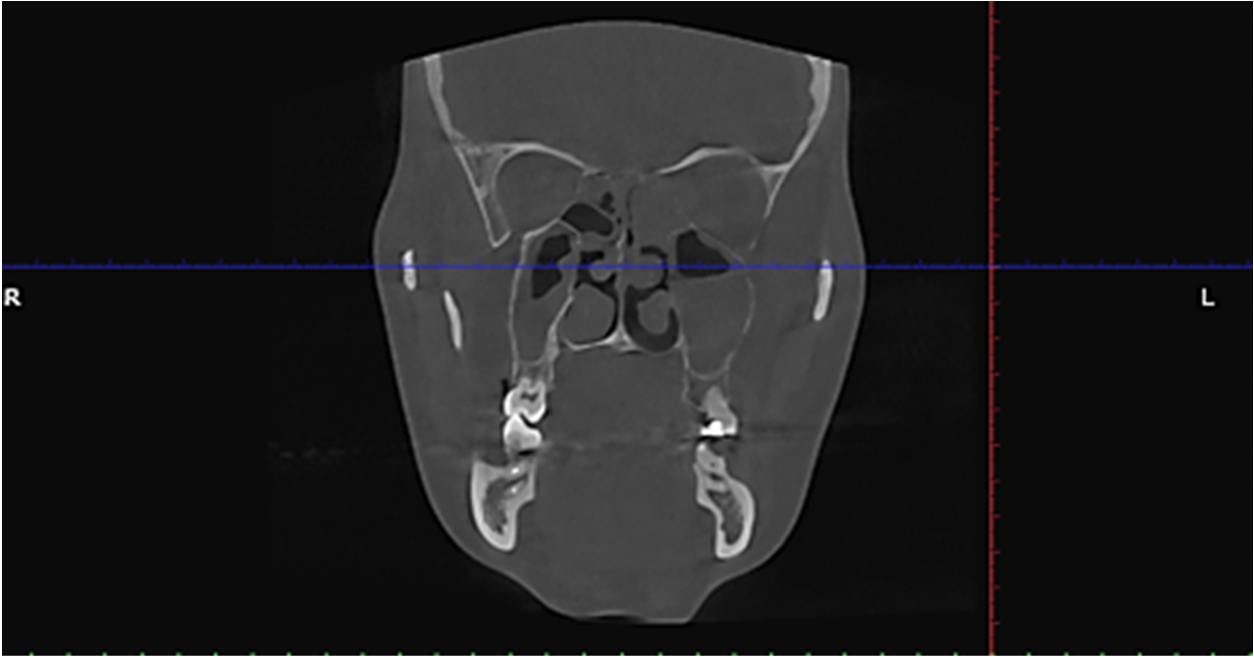
Морфологічне дослідження біопсійного матеріалу. Видалений під час хірургічного втручання матеріал фіксовано в розчині 10% нейтрального забуференого формаліну та доставлено в патологоанатомічне відділення, де тканини було зневоднено, знежирено та занурено в парафін для проведення гістологічного дослідження. З отриманих парафінових блоків з тканиною виготовлено тонкі зрізи товщиною 4 мкм. Після депарафінізації та регідратації зрізи забарвлено за стандартною методикою гематоксиліном й еозином та вміщено під покривне скло за допомогою монтажного середовища на основі ксилолу. Цифрові фотографії отримано за допомогою кольорової камери мікроскопа Zeіss Prіmo Star – Axіocam ERC 5s з ліцензійним програмним забезпеченням ZEN 2 blue edіtіon (збільшення ×100/400). При гістологічному дослідженні матеріалу, вилученого з гайморової пазухи, серед некротичного детриту, на тлі щільного запального інфільтрату визначалася велика кількість гранульом, часто зливних, які за своєю морфологією відповідали актиномікотичним (рис. 3). У центрі гранульом виявлено базофільні утворення округлої або овоїдної форми, які представляли собою конгломерати радіально розташованих тонких, нитчастих, грибоподібних мікроорганізмів з вираженим розгалуженням («сірчані гранули»), оточені вузьким обідком еозинофільної аморфної речовини (рис. 4). Запальний інфільтрат був неоднорідним та складався переважно з мононуклеарних клітин (лімфоцитів, плазмоцитів, моноцитів), серед яких також знаходилися чисельні нейтрофіли різного ступеня зрілості, поодинокі еозинофіли та макрофаги (рис. 5), що свідчило про наявність хронічного активного запалення з тенденцією до нагноєння.
Рис. 3. Множинні зливні актиномікотичні гранульоми серед некротичного детриту (Г+Е, ×100): 1 – «сірчані гранули»; 2 – гетерогенний запальний інфільтрат; 3 – детрит з окремими бактеріальними філаментами ↓
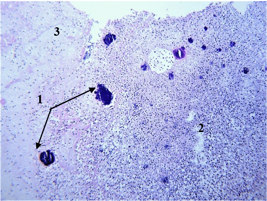
Рис. 4. Типова «сірчана гранула» серед запального інфільтрату та некротичного детриту (Г+Е, ×400): 1 – базофільний конгломерат нитчастих мікроорганізмів; 2 – еозинофільний аморфний обідок; 3 – нейтрофіли; 4 – детрит ↓
Рис. 5. Гетерогенний запальний інфільтрат навколо «сірчаних гранул» з лімфоцитів, плазмоцитів, моноцитів, нейтрофілів, а також поодиноких еозинофілів та макрофагів (Г+Е, ×400) ↓
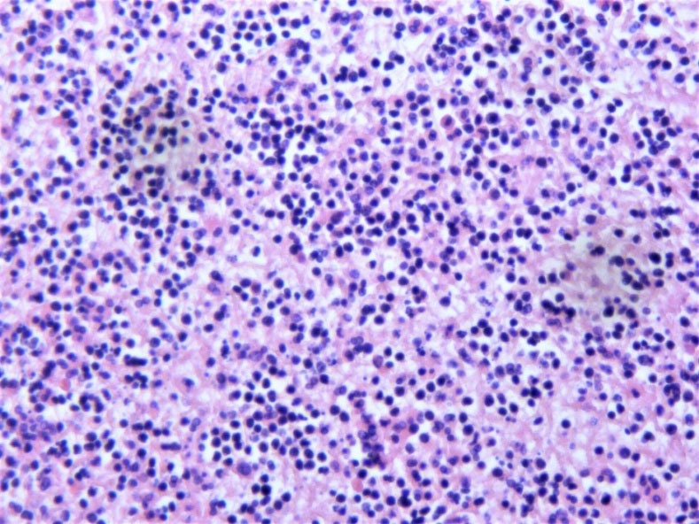
25.12.2024 року пацієнтка направлена на консультацію до лікаря-інфекціоніста. Рішенням консиліуму в складі співробітників кафедри інфекційних хвороб ДДМУ було призначено внутрішньовенно цефтріаксон 2,0 г 1 раз на добу. Тривалість та ефективність лікування планувалось оцінювати за клінічним перебігом, насамперед станом гайморових пазух, відсутністю несприятливих реакції, а також мікробіологічним контролем, але не менше ніж 28 днів. Ураховуючи стабільний, задовільний клінічний статус, задля зменшення ризику госпітального інфікування, лікування проводилось амбулаторно, було складено графік відвідувань лікаря-оториноларинголога та інфекціоніста. 06.01.2025 пацієнтка раптово втратила свідомість, у зв’язку з чим була госпіталізована у відділення інтенсивної терапії обласної лікарні. При КТ-дослідженні головного мозку – виключені пухлинна та судинна патологія, при дослідження спинномозкової рідини – показники без патологічних змін. Призначений моксифлоксацин 0,4 г 1 раз на добу, внутрішньовенно тривалістю 7 днів. При повторному огляді лікаря-оториноларинголога – ураховуючи відсутність ознак запалення та новоутворення при ревізії носової порожнини, а також задовільний загальний клінічний статус було ухвалено рішення не відновлювати системну антибактеріальну хіміотерапію. Таким чином, наша пацієнтка отримала двоетапний курс системної антибактеріальної терапії загальною тривалістю 19 днів, який привів до санації носової порожнини. На цей час пацієнтка має задовільний стан, регулярно обстежується оториноларингологом, спостерігається лікарями інфекціоністом та ендокринологом.
РЕЗУЛЬТАТИ ТА ЇХ ОБГОВОРЕННЯ
Цей клінічний випадок актиномікозу носових пазух поставив перед нами ряд викликів клінічного, організаційного та особистого плану. Проблемою, яка виникла, була складність виконати пряму верифікацію збудника класичним (культуральним) методом або шляхом ідентифікації ДНК актиноміцет методом ПЛР, що певною мірою обумовлено обмеженням можливості культивувати анаероби, які є найбільш вірулентними, та визначати вид Actinomyces. Лабораторна верифікація інфекції Actinomyces на підставі морфологічних досліджень достатньо суб’єктивна і залежить від кваліфікації морфолога, тому маючи тільки такий лабораторний метод, секційний матеріал було перевірено двома незалежними патогістологічними лабораторіями. За висновком морфологічного дослідження секційний матеріал містив актиноміцети. Водночас Actinomyces spp. є представниками нормальної мікрофлори слизових оболонок людини, тому його виділення само по собі не є доказом, що саме цей збудник є причиною захворювання. Доказом причинності Actinomyces spp. є його виділення з вогнища ураження, що, стосовно патології ЛОР-органів, вимагає від лікаря певних навичок і кваліфікації (досвіду) у відборі матеріалу, його зберіганні і транспортуванні до лабораторії, щоб уникнути зовнішньої контамінації біопсійного зразка. Слід також звернути увагу на можливі умови, що сприяли захворюванню на актиномікоз у жінки без порушень імунітету: цукровий діабет, інвазивні стоматологічні та отоларингологічні процедури й нераціональну тривалу системну антибактеріальну хіміотерапію, яка могла призвести до дисбіозу слизової оболонки пазух та, відповідно, до активації коменсалів. Зазначені можливі фактори ризику збігаються з даними інших спостережень [9]. У зв’язку з нераціонально призначеною тривалою антибактеріальною терапією, постало питання − хто і як повинен лікувати цю пацієнтку. Враховуючи різну топіку можливих клінічних проявів Actinomyces spp., ведення пацієнтів з цією інфекцію має здійснювати мультидисциплінарна команда в такому складі: інфекціоніст, за топікою ураження − відповідний фахівець, клінічний мікробіолог, патоморфолог, якщо хворобу діагностовано в дитини − педіатр. Згідно з наявними результатами спостережень [2, 9], оптимальним лікуванням актиномікозу є довготривала антибактеріальна терапія, зокрема, як у нашому випадку – призначення щоденного внутрішньовенного введення цефтріаксону на 28 днів.
Оскільки перебіг актиномікозу є варіативним ‒ з розвитком або деструктивної форми з утворенням абсцесів, або деструктивно-проліферативної форми захворювання, а в рідкісних випадках ‒ з відсутністю специфічних макроскопічних змін в уражених тканинах, що може ускладнювати діагностичний процес [4], існує необхідність ретельного дослідження операційного матеріалу із застосуванням гістологічного методу [10, 11]. Зазвичай для верифікації діагнозу достатньо рутинного забарвлення зрізів тканин гематоксиліном й еозином, однак інколи також застосовують забарвлення за Цілем-Нільсеном або Кіньюном [12]. Типовою мікроскопічною ознакою актиномікозу є наявність специфічних гранульом з центрально розташованими округлими або неправильної форми базофільними агрегатами ниткоподібних мікроорганізмів ‒ «сірчаними гранулами», оточеними еозинофільним безструктурним матеріалом та різнорідним валом клітин запального ряду [13].
Амбулаторне лікування таких пацієнтів може зменшити ризики госпітальної інфекції і навантаження на систему громадського здоров’я, а також уникнути соціальної дезадаптації. Водночас при тривалому амбулаторному лікуванні можуть виникнути екстремальні ситуації, що здатні суттєво вплинути або звести нанівець результат лікування, як у нашому випадку. Важливим рішенням цієї проблеми є постійна комунікація з лікарем та обговорення заздалегідь з пацієнтом плану дій у виникненні надзвичайних ситуацій. Відсутність особистого практичного досвіду в лікуванні пацієнтів з подібної патологією уповільнило діагностику та призначення лікування відповідної тривалості.
ВИСНОВКИ
1. Хронічні, тривалі або рецидивуючі гранулематозні ураження ротової порожнини та оториноларингологічних органів, які не «відповідають» на стандартну антибактеріальну терапію, потребують ретельної диференційної діагностики та виключення Actinomyces spp. − етіології хвороби.
2. Актиномікоз у такому випадку був верифікований за допомогою гістологічного методу з ретельним дослідженням операційного матеріалу, що дозволило відкоригувати лікування.
3. Підвищення можливостей мікробіологічних лабораторій щодо виділення та ідентифікації анаеробів, зокрема Actinomyces spp., є важливим фактором для вирішення проблеми діагностики та лікування цієї інфекції.
4. Діагностика й лікування актиномікозу потребувало мультипрофесійної участі, у нашому випадку були задіяні інфекціоністи, отоларинголог, патоморфолог.
5. Необхідним для клінічної практики є створення відповідних клінічних настанов щодо діагностики та лікування рідкісних інфекційних хвороб, зокрема актиномікозу.
Внески авторів:
Литвин К.Ю. – дослідження, ідея, написання-редагування, рецензування;
Усова О.М. – патоморфологічне дослідження, написання-редагування;
Білоконь О.О. – візуалізація, формальний аналіз, написання-редагування;
Шостакович-Корецька Л.Р. – рецензування;
Маврутенков В.В. – методологія, курація даних, написання – початковий проєкт;
Гутников П.В. – курація даних, візуалізація, написання;
Волікова О.О. – технічне редагування.
Фінансування. Дослідження не має зовнішніх джерел фінансування.
Конфлікт інтересів. Автори заявляють про відсутність конфлікту інтересів.
REFERENCES
1. Moniruddin A, Begum H, Nahar K. Actinomycosis: an update. Med Today. 2012 Nov 4;21(2):38-42. doi: https://doi.org/10.3329/medtoday.v21i2.12478
2. Boyanova L, Kolarov R, Mateva L, Markovska R, Mitov I. Actinomycosis: a frequently forgotten disease. Future Microbiology. 2015;10(4):613-28. doi: https://doi.org/10.2217/fmb.14.130
3. De Simeis D, Serra S. Actinomycetes: A Never-Ending Source of Bioactive Compounds-An Overview on Antibiotics Production. Antibiotics (Basel). 2021;10(5):483. doi: https://doi.org/10.3390/antibiotics10050483
4. Cohn JE, Lentner M, Li H, Nagorsky M. Unilateral maxillary sinus actinomycosis with a closed oroantral fistula. Case Reports in Otolaryngology. 2017;2017:1-3. doi: https://doi.org/10.1155/2017/7568390
5. Rothschild B, Naples V, Barbian L. Bone manifestations of actinomycosis. Annals of Diagnostic Pathology. 2006;10(1):24-7. doi: https://doi.org/10.1016/j.anndiagpath.2005.09.017
6. Wadhera R, Gulati SP, Garg A, Ghai A, Kumar S. Frontal sinus actinomycosis presenting as osteomyelitis of frontal bone. Otolaryngology – Head and Neck Surgery. 2008;138(4):544-5. doi: https://doi.org/10.1016/j.otohns.2007.12.009
7. Woo H-J, Hoon BC, Song Si-Youn, et al. Actinomycosis of the paranasal sinus. Otolaryngology – Head and Neck Surgery. 2008;139(3):460-2. doi: https://doi.org/10.1016/j.otohns.2008.06.001
8. Varghese L, Cherian LM, Varghese GM. Actinomycosis: An Unusual Cause of Maxillary Sinusitis. Ear, Nose & Throat Journal. 2020;101(7):014556132096520. doi: https://doi.org/10.1177/0145561320965207
9. Valour F, Sénéchal A, Dupieux C, Karsenty J, Lustig S, Breton P, et al. Actinomycosis: etiology, clinical features, diagnosis, treatment, and management. Infect Drug Resist. 2014 Jul 5;7:183-97. doi: https://doi.org/10.2147/idr.s39601
10. Roychowdhury D, Chatterjee RP, Gayen S, Das S, Chatterjee A, Bagchi S, et al. Oral Granulomatous Disorders: A Diagnostic Insight. Cureus. 2024 Jul 30;16(7):e65742. doi: https://doi.org/10.7759/cureus.65742
11. Terashima K, Kagoya R, Tanaka M, Nishijima H, Kondo K. A case of coexistent fungal rhinosinusitis and actinomycosis caused by Gutta-Percha points extruded into the maxillary sinus. Cureus. 2024 Sep 26;16(9):e70246. doi: https://doi.org/10.7759/cureus.70246
12. Pérez-Sayáns M, Suárez Peñaranda JM, Quintanilla JAS, et al. Clinicopathological features of 214 maxillary sinus pathologies. A ten-year single-centre retrospective clinical study. Head Face Med. 2020;16(1):24. doi: https://doi.org/10.1186/s13005-020-00239-x
13. Arshad W, Mahmood Kamal M, Rafique Z, Rahat M, Mumtaz H. Case of maxillary actinomycotic osteomyelitis, a rare post COVID complication-case report. Ann Med Surg (Lond). 2022;80:104242. doi: https://doi.org/10.1016/j.amsu.2022.104242
 UK
UK  EN
EN